Translate this page into:
Establishing a protocol for immunocytochemical staining and chromogenic in situ hybridization of Giemsa and Diff-Quick prestained cytological smears
*Corresponding author
-
Received: ,
Accepted: ,
This is an open-access article distributed under the terms of the Creative Commons Attribution-Noncommercial-Share Alike 3.0 Unported, which permits unrestricted use, distribution, and reproduction in any medium, provided the original work is properly cited.
This article was originally published by Medknow Publications & Media Pvt Ltd and was migrated to Scientific Scholar after the change of Publisher.
Abstract
Background:
Protocols for immunocytochemical staining (ICC) and in situ hybridization (ISH) of air-dried Diff-Quick or May-Grünwald Giemsa (MGG)-stained smears have been difficult to establish. An increasing need to be able to use prestained slides for ICC and ISH in specific cases led to this study, aiming at finding a robust protocol for both methods.
Materials and Methods:
The material consisted of MGG- and Diff-Quick-stained smears. After diagnosis, one to two diagnostic smears were stored in the department. Any additional smear(s) containing diagnostic material were used for this study. The majority were fine needle aspirates (FNAC) from the breast, comprising materials from fibroadenomas, fibrocystic disease, and carcinomas. A few were metastatic lesions (carcinomas and malignant melanomas). There were 64 prestained smears. Ten smears were Diff-Quick stained, and 54 were MGG stained. The antibodies used for testing ICC were Ki-67, ER, and PgR, CK MNF116 (pancytokeratin) and E-cadherin. HER-2 Dual SISH was used to test ISH. Citrate, TRS, and TE buffers at pH6 and pH9 were tested, as well as, different heating times, microwave powers and antibody concentrations. The ICC was done on the Dako Autostainer (Dako®, Glostrup, Denmark), and HER-2 Dual SISH was done on the Ventana XT-machine (Ventana / Roche® , Strasbourg, France).
Results:
Optimal results were obtained with the TE buffer at pH 9, for both ICC and ISH. Antibody concentrations generally had to be higher than in the immunohistochemistry (IHC). The optimal microwave heat treatment included an initial high power boiling followed by low power boiling. No post fixation was necessary for ICC, whereas, 20 minutes post fixation in formalin (4%) was necessary for ISH.
Conclusions:
Microwave heat treatment, with initial boiling at high power followed by boiling at low power and TE buffer at pH 9 were the key steps in the procedure. Antibody concentrations has to be adapted for each ICC marker. Post fixation in formalin is necessary for ISH.
Keywords
Immunocytochemical
in situ hybridization
MGG
prestained
Tris-EDTA
INTRODUCTION
Cytological investigation is a valuable first option in the workup of a suspected tumor, practically anywhere in the body. FNAC is a fast, simple, and cheap procedure that usually does not require a local anesthetic in superficial settings. A preliminary diagnosis, ‘on the spot’ (ROSE = rapid on site evaluation),[1–6] is rendered possible by this method.
There is an increasing demand for more specific diagnoses, and a diagnosis of ‘malignant cells’ or ‘carcinoma’ is in many instances not enough to determine the optimal primary management of the patient. Thus follows an increasing need for subtyping of tumors and for analysis of prognostic, predictive, and therapeutic markers, prior to the eventual surgery or preoperative chemotherapy. Both immunocytochemistry (ICC) and in situ hybridization (ISH) is increasingly being done on cytological preparations, both on direct smears, cell blocks, and liquid-based preparations.[7–13] In larger institutions it is common that cytopathologists or cytotechnologists attend when a suspicious lesion is being sampled for diagnostic purposes. Additional material for ICC or ISH may then be obtained whenever needed. For a variety of reasons, personnel from the Pathology Department may not be present, and the laboratories receive alcohol-fixed or air-dried unfixed smears for diagnosis. All the received smears are usually stained for a primary diagnostic workup. ICC on Papanicolaou-stained materials is usually possible.[14–17] However, it has turned out to be quite difficult to do ICC on May Grunwaald Giemsa (MGG) or Diff-Quick® prestained smears. Recently, an article from Choi et al.[18] has described a protocol for Ki-67 staining on Diff-Quick-stained smears from breast tumors in dogs. The aim of this study is to develop a robust protocol for ICC and ISH, which could work on air-dried MGG- and Diff-Quick-stained smears.
MATERIALS AND METHODS
The material consisted of MGG- and Diff-Quick-stained, direct smears. After diagnosis, one to two diagnostic smears were stored in the department and any additional smear(s) containing diagnostic material were used for this study. The vast majority were fine needle aspirates (FNAC) from the breast, comprising materials from fibroadenomas, fibrocystic disease, and carcinomas. A few were metastatic lesions (carcinomas and malignant melanomas). There were 64 prestained smears, 10 of them were Diff-Quick stained and 54 were MGG. The primary material was collected consecutively for two months. Adjunct diagnostic slides from cases were chosen to mimic the setting in which this method would be used. The slides were only used once and subsequent staining on negative test slides was not done. Smears and antibodies were matched according to the morphological, cytological, and (eventually also) histological diagnosis. In doing so, we knew that the tumor cells should be positive. Negative ICC test stains were then per se false negative. The non-epithelial cells (lymphocytes and stromal cells) served as the internal negative controls for all markers except Ki-67. Separate negative and positive controls were not used, but are essential in a diagnostic setting.
The optimal procedure was retested for all five antibodies. The HER-2 Dual SISH probe was retested twice. In addition, the ICC procedure was tested twice in our routine cytology ICC laboratory. These last smears (n = 10 breast carcinomas) were collected three months later in the same manner as stated earlier.
The antibodies used for testing ICC were Ki-67 (Dako®, Glostrup, Denmark), ER (Novocastra Laboratories®, Newcastle, UK), and PgR (Novocastra Laboratories®, Newcastle, UK), as representatives of nuclear epitopes, a pancytokeratin (CK MNF116, Dako®, Glostrup, Denmark) for intracytoplasmic markers, and E-cadherin (Dako®, Glostrup, Denmark) as a representative of a cytoplasmic membrane epitope. HER-2 Dual SISH (Ventana INFORM HER2 Dual Color ISH, Roche®, Strasbourg, France) was used to test ISH.
Different types of buffer (Citrate, TE, TRS) and pH (6 vs. 9) were tested, as well as different heating times and microwave powers, as also the concentration of the antibodies.
The smears were put in xylene to remove the coverslips. Rehydration was done using 100, 96, and 70% ethanol. Formalin was used for post fixation when testing ISH. No post fixation was used for ICC. The ICC was done on the Dako Autostainer (Dako®, Glostrup, Denmark) and HER-2 Dual SISH was done on the Ventana XT-machine (Ventana / Roche®, Strasbourg, France).
RESULTS
Some extraction of the stain from the slides was noticed during rehydration, seen as a light blue discoloration of the jar containing 70% ethanol. Details of the ICC results are shown in Table 1. Boiling Ki-67 stained slides in citrate buffer in MW for 2 × 5 minutes at high power (Choi et al.), worked, but was suboptimal. For the other nuclear markers (ER and PgR) this protocol was unsuccessful. We tested the same buffer with different boiling times and microwave powers for all the other antibodies. There was no destaining, and ICC was negative. The TRS buffer (pH 6.0) was tested for different boiling times and microwave powers, with negative results. Citrate and TRS buffers are used for some primary antibodies in Heat-Induced Epitop Retrieval (HIER) pretreatment, but were unsuitable in our testing. Both buffers have a pH of 6. The TE-buffer set at high microwave power for only 2.5 minutes and then lower power to keep it boiling for six minutes was efficient for destaining and pretreatment for all three nuclear markers. The same was true for the intracytoplasmic marker Cytokeratin MNF116 and the membrane marker E-Cadherin. When tested in a routine laboratory with a different microwave oven, three-and-a-half minutes followed by seven minutes at low power, were necessary to give the equivalent results. The protocol turned out to be equal for epitopes in all three cellular compartments (nuclear, cytoplasmic, and membranic) and is shown in Table 2.
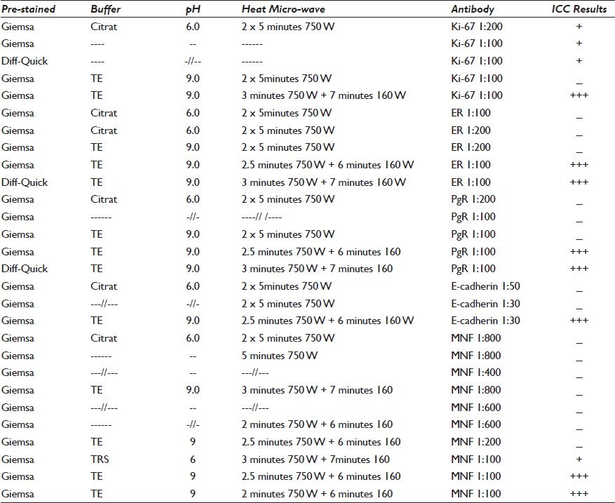

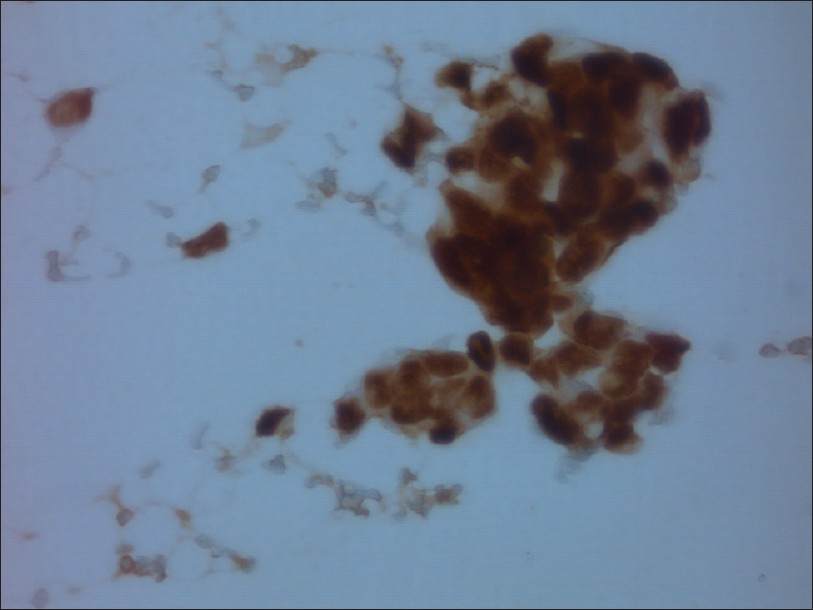
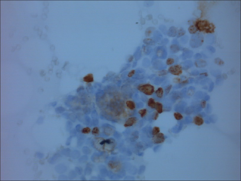
The ICC results for all tested antibodies showed a good and easily interpretable specific staining [Figures 1–5]. There was almost no background staining. In some cases, the background staining was interpreted as staining of cytoplasmic content from disrupted cells (Cytokeratin MNF116 and E-cadherin) and thus as probably ‘specific’. The same TE buffer, at only a high power of 750 W, for a prolonged time (2 × 5 minutes) of microwave treatment gave negative results for ER and PgR and weak positivity for Ki-67.
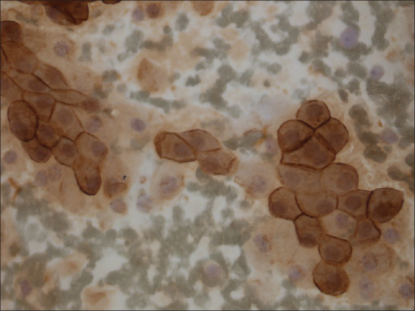
- E-cadherin staining of cells from ductal breast carcinoma (Magnification, ×400)
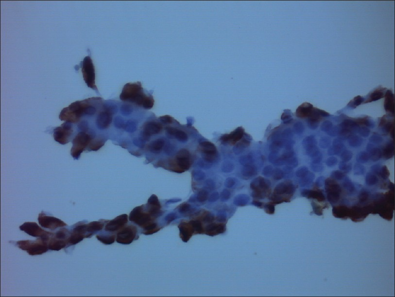
- Cytokeratin MNF116 (pancytokeratin) staining of benign apocrine cells from fibrocystic disease (Magnification, ×400)
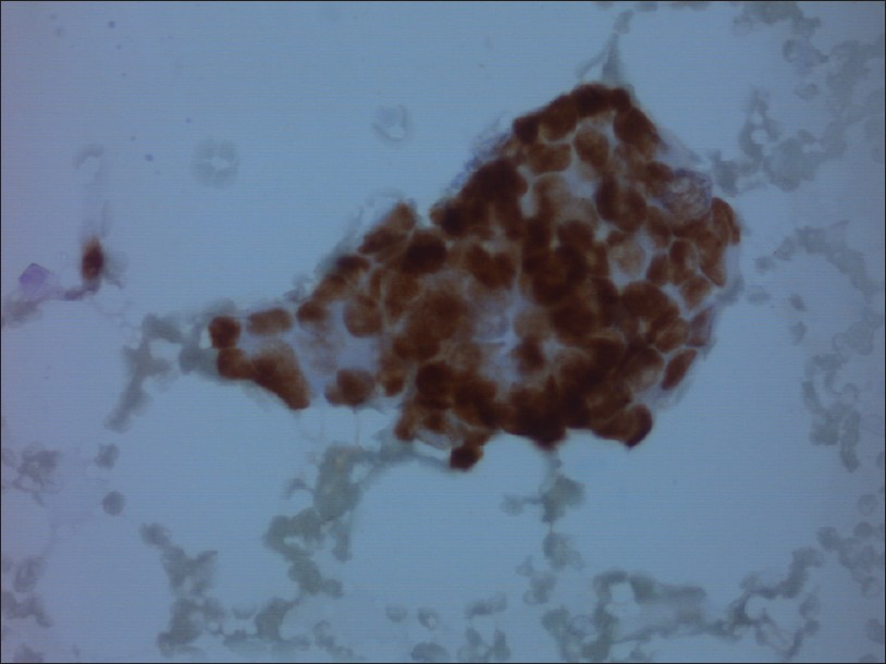
- ER positive, benign ductal cells (Magnification, ×400)
- PgR positive, benign ductal cells (Magnification, ×400)
- Ki-67 positive nuclei from ductal breast carcinoma (Magnification, ×400)
For all ICC markers we had to increase the antibody concentration compared to the concentration used in the routine practice of ICC and / or immunohistochemistry (IHC). The concentration of Cytokeratin MNF116 turned out to be remarkably higher, whereas, the other antibodies needed a somewhat higher concentration.
The test results for HER-2 Dual SISH are shown in Table 3. The signal numbers and HER-2 status were identical to the results in the routine histological examination. The best signal intensity of both the red and silver stains was obtained with 20 minutes post fixation in formalin and TE buffer, with pH 9 [Figure 6]. The specific hybridization steps were not changed. Here also, the details of the microwave treatment should be optimized locally.

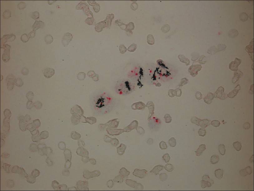
- Dual SISH HER-2 from metastatic breast carcinoma. The tumor cells are amplified with large clusters of HER-2 silver signals and 2 (red) signals of chromosome 17 (CEP17). One lymphocyte reveals two signals of both the HER-2 gene and CEP17 (Magnification, ×400)
DISCUSSION
Our objective was to find out how to manipulate or break down the stain-cell attractive forces made by the MGG and Diff-Quick stain-cell affinities. The origins of stain-cell affinity (the tendency of a stain to move from the staining solution into the cell) make it possible for the cells to stain. Cytological stains like the Papanicolaou (PAP) and Romanowsky-Giemsa methods involve mixtures of acid and basic dyes. By Coulomb`s law, electric forces arise between these ionic stains and the cellular constituents (PO 4- , SO4 - , COO -, NH3 +) of opposite electric charges. Destaining PAP-stained smears in a solution containing HCL, ethanol, and H2O with antigen recovery is efficient. The Romanowsky-Giemsa staining involves a more complicated chemistry and so does the destaining. First, the cation dye (methylene blue derivative Azure B) and anion dye (Eosin Y) are in the same solution. Second, the purple (metachromatic) stain of the nuclei is believed to be due to the formation of a stain-stain attraction of the methylene blue derivative Azure B and Eosin Y complex to the cell chromatin.[1920] Some destaining was evident during the last step of rehydration (70% ethanol). This may support the theory that water is slightly basic and thus extracts the acid (anion) dye. It might also be that the cells swell and the dyes aggregate more in aqueous solution than in pure alcohol.
As can be seen in Table 1, the type of heating buffer and its pH had a significant effect on slide destaining and in the further antigen-antibody interaction on the ICC. Boiling slides in a microwave oven with Tris-EDTA buffer (TE-buffer), pH 9.0, gave successful ICC and ISH restaining. The TE-buffer has triple advantages. First, we could assume that the high pH (higher than 6.5 in the MGG stain) and the high salt content of this buffer makes the staining bonds weaker. The negative and positive ions in the buffer could compete with the respective negative- and positive-charged dye molecules to bind to the cell components. The dye molecules might wholly or in part be ousted and lose their binding site with the cell components (PO4-, SO4-, COO-, NH3+).[21] It is known that dyes have a tendency to aggregate in aqueous salty solutions, and this could lead to a reduced reactivity.[2223] Other possible factors that contributed to dye aggregation could include the acid-base properties, hydrophobic interactions, possibly interference of metal-complex formation, van der Waals′ forces, and the entropy effects.
There are two features that make Ethylenediaminetetraacetic acid (EDTA) a masking agent. It forms stable, water-soluble, chelate complexes with most metal ions. Its complexing ability is pH-dependent and EDTA is a strong ligand at high pH.[24] According to Wittekind,[25] azure B and eosin are associated in part, due to their hydrophobic effects, which may explain the lability of the complex when exposed to solvents, which disrupt the water structure or remove the water completely.[25–27] In the case of the MGG or Diff-Quick stain, the EDTA's chelating effect may replace the water molecules.
The third issue is microwave heating. The greater the energy supplied to the system, that is, the higher the temperature, the weaker the binding force will be. TE-buffer pH 9.0 is the antigen retrieval buffer of choice for many primary antibodies that require pretreatment. Determination of the buffer type with a respective boiling time and microwave power has been made after comparative tests with different variables. TE-buffer pH 9.0 with a short boiling time (2.5 minutes) at high power (750 W), with further boiling at a lower power (160 W), for six minutes, gave good positive ICC staining and clear ISH signals. Different microwave ovens may require modifications to this.
Antibody concentrations were generally higher than in our routine procedures and should be established for each antibody used. Positive ICC results were reliable as to being positive or negative, but the staining intensity was generally lower. The percentage of positive nuclei was lower than in the routine specimens and known / established cut-off values for Ki-67, ER or PgR cannot be used. Caution should be exercised as to false positive results. When the antibody titer is very high, excessive inflammation in the background, marked necrosis, and crushed and degenerated cells could cause false positive staining.
Proper positive and negative controls must always be applied in a diagnostic setting. In this study we have focused on establishing a protocol for staining in cases where we knew the tumor cells should be positive and thus they were their own positive controls. Internal non-epithelial cells such as lymphocytes and stromal cells served as the negative control. Depending on the availability of fresh material it would be feasible to make smears, stain with Diff-Quick or Giemsa, and use these as positive and negative controls. Another alternative is to store diagnostic slides with known immunoprofiles and used them as positive and negative controls.
An advantage of using prestained FNA smears or other cytological materials is the ability to identify a slide with adequate or abundant diagnostic cells. The use of archival cytological slides to perform ISH with specific genes and / or chromosomes and ICC status is economical, practical, and feasible, when unstained smears, liquid-based material or tissue sections are not available.
In conclusion, initial microwave boiling at high power followed by further boiling at low power, using TE buffer at pH 9, seem to be the crucial features for successful ICC and ISH on Diff-Quick and MGG prestained smears. Optimal antibody concentrations and optimal times for microwave treatment must be determined in each laboratory. By means of the protocol described here, it is possible to carry out sequential (re)-staining of different antibodies and ISH probes.
COMPETING INTEREST STATEMENT BY ALL AUTHORS
There is no competing interest to declare by any of the authors.
AUTHORSHIP STATEMENT BY ALL AUTHORS
Elsa Beraki has designed the protocols for testing and their modifications, done the literature search, and most of the primary writing of the manuscript. Thale Kristin Olsen did the practical work, literature search and most of the introduction of the manuscript. Torill Sauer was the senior researcher responsible for the idea and for the submitted version of the manuscript.
ETHICS STATEMENT BY ALL AUTHORS
This study was conducted with the approval from the Institutional Review Boards (IRB) (or its equivalent) of all the institutions associated with this study, as applicable. Authors take responsibility to maintain relevant documentation in this respect.
EDITORIAL / PEER-REVIEW STATEMENT
To ensure the integrity and highest quality of CytoJournal publications, the review process of this manuscript was conducted under a double blind model (authors are blinded for reviewers and vice versa) through automatic online system.
ACKNOWLEDGMENTS
Each author acknowledges that this final version was read and approved. All authors declare that they qualify for authorship as defined by ICMJE http:///www.icmje.org/#author. Each author has participated sufficiently in the study and takes public responsibility for the appropriate portions of the content in this article.
Available FREE in open access from: http://www.cytojournal.com/text.asp?2012/9/1/8/94518.
REFERENCES
- Immediate interpretation of FNA smears from the head and neck region. Diagn Cytopathol. 1992;8:116-8.
- [Google Scholar]
- Utility of rapid on-site evaluation of transbronchial needle aspirates. Respiration. 2005;72:182-8.
- [Google Scholar]
- Impact of rapid on-site cytologic evaluation during transbronchial needle aspiration. Chest. 2005;128:869-75.
- [Google Scholar]
- Endoscopic ultrasound-guided fine-needle aspiration of solid pancreatic masses with rapid on-site cytological evaluation by endosonographers without attendance of cytopathologists. J Gastroenterol. 2009;44:322-8.
- [Google Scholar]
- Role and accuracy of rapid on-site evaluation of CT-guided fine needle aspiration cytology of lung nodules. Cytopathology. 2011;22:306-12.
- [Google Scholar]
- Comparison of the quality of smears in transbronchial fine-needle aspirates using two staining methods for rapid on-site evaluation. Diagn Cytopathol 2011 [In Press]
- [Google Scholar]
- Control specimens for immunocytochemistry in liquid-based cytology. Cytopathology. 2011;22:243-6.
- [Google Scholar]
- External quality control for immunocytochemistry on cytology samples: a review of UK NEQAS ICC (cytology module) results. Cytopathology. 2011;22:230-7.
- [Google Scholar]
- Immunocytochemistry in Europe: results of the European Federation of Cytology Societies (EFCS) inquiry. Cytopathology. 2011;22:238-42.
- [Google Scholar]
- Liquid based material from fine needle aspirates from breast carcinomas offers the possibility of long-time storage without significant loss of immunoreactivity of estrogen and progesterone receptors. Cytojournal. 2010;7:24.
- [Google Scholar]
- Determination of HER-2 status on FNAC material from breast carcinomas using in situ hybridization with dual chromogen visualization with silver enhancement (dual SISH) Cytojournal. 2010;7:21.
- [Google Scholar]
- Assessing estrogen and progesterone receptor status in fine needle aspirates from breast carcinomas.Results on six years of material and correlation with biochemical assay. Anal Quant Cytol Histol. 1998;20:122-6.
- [Google Scholar]
- Estrogen and progesterone receptor status in breast carcinoma: comparison of immunocytochemistry and immunohistochemistry. Diagn Cytopathol. 2002;26:137-41.
- [Google Scholar]
- Immunocytochemical evaluation of estrogen receptor on archival Papanicolaou-stained fine-needle aspirate smears. Diagn Cytopathol. 2003;29:309-14.
- [Google Scholar]
- MIB-1 immunostaining on cytological samples: a protocol without antigen retrieval. Cytopathology. 2004;15:154-9.
- [Google Scholar]
- Efficacy of Ki-67 antigen staining in Papanicolaou (Pap) smears in post-menopausal women with atypia--an audit. Cytopathology. 1999;10:369-74.
- [Google Scholar]
- Usefulness of estrogen receptor detection using archival Papanicolaou-stained smears. Acta Cytol. 1999;43:825-30.
- [Google Scholar]
- Immunocytochemical detection of Ki-67 in Diff-Quik-stained cytological smears of canine mammary gland tumours. Cytopathology. 2011;22:115-20.
- [Google Scholar]
- Understanding Romanowsky staining.I: The Romanowsky-Giemsa effect in blood smears. Histochemistry. 1987;86:331-6.
- [Google Scholar]
- The interaction of methylene blue, azure B, and thionine with DNA: Formation of complexes with polynucleotides and mononucleotides as model systems. Biopolymers. 1995;35:419-33.
- [Google Scholar]
- The interrelation of the size and substantivity of dyes; the role of van der Waals attractions and hydrophobic bonding in biological staining. Histochemie. 1973;33:191-204.
- [Google Scholar]
- Some effects of salts on staining: use of the Donnan equilibrium to describe staining of tissue sections with acid and basic dyes. Histochemistry. 1974;39:71-82.
- [Google Scholar]
- (1969), Aggregation of Dyes in Aqueous Solutions. Journal of the Society of Dyers and Colourists. 0;85:355-368. doi: 101111/j1478-44081969tb02909x
- [Google Scholar]
- Masking and demasking of chemical reactions: theoretical aspects and practical applications. Hoboken, New Jersey: Wiley-Interscience; 1970.
- [Google Scholar]
- On the nature of Romanowsky-Giemsa staining and the Romanowsky-Giemsa effect. I. Model experiments on the specificity of azure B-eosin Y stain as compared with other thiazine dye-eosin Y combinations. Histochem J. 1985;17:263-89.
- [Google Scholar]
- How Romanowsky stains work and why they remain valuable - including a proposed universal Romanowsky staining mechanism and a rational troubleshooting scheme. Biotech Histochem. 2011;86:36-51.
- [Google Scholar]
- A critical evaluation of Bernhard’s EDTA regressive staining technique for RNA. J Cell Sci. 1982;54:207-40.
- [Google Scholar]


