Translate this page into:
Lung tumor in a young African American patient with sickle trait: Pieces of a puzzle
*Corresponding author
-
Received: ,
Accepted: ,
This is an open access journal, and articles are distributed under the terms of the Creative Commons Attribution-NonCommercial-ShareAlike 4.0 License, which allows others to remix, tweak, and build upon the work non-commercially, as long as appropriate credit is given and the new creations are licensed under the identical terms.
This article was originally published by Medknow Publications & Media Pvt Ltd and was migrated to Scientific Scholar after the change of Publisher.
PERTINENT CLINICAL HISTORY TO GUIDE THE QUIZ
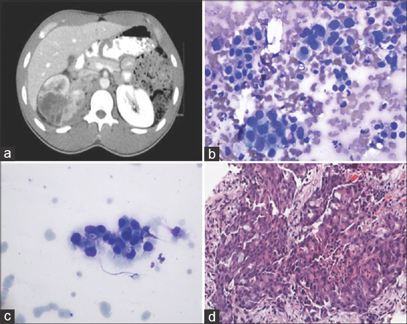
A 32-year-old African American male with sickle cell trait presented with headaches, nausea, vomiting, cough, pleuritic chest pain, hypertension, and a 10-pound weight loss. A computed tomography (CT) showed multiple lung nodules, mediastinal lymphadenopathy, and an 11 cm mass in the right kidney with a suspicion for a tumor thrombus in the renal vein. Figure 1a–d shows an abdominal CT, cytomorphological features, and an hematoxylin and eosin (H and E) of the lung core biopsy.

- (a) Computed tomography scan of the abdomen showing an 11 cm right renal mass. (b and c) Touch imprints of the lung core biopsy showing malignant cells arranged singly and in loose clusters with large, hyperchromatic nuclei, irregular nuclear contours and a moderate amount of granular cytoplasm with dense eosinophilic cytoplasmic globules (Diff-Quik stain, ×400). (d) Core biopsy showing malignant cells arranged in nests with areas of cribriform pattern and acute inflammation in a background of stromal fibrosis. Many cells demonstrate rhabdoid features. (H and E, ×400)
WHAT IS YOUR INTERPRETATION?
-
Metastatic clear cell renal cell carcinoma (RCC)
-
Primary lung non small cell carcinoma
-
Metastatic renal medullary carcinoma (RMC)
-
Metastatic high-grade urothelial carcinoma.
ANSWER
The correct cytopathologic interpretation is:
c. Metastatic renal medullary carcinoma (RMC)
BRIEF DISCUSSION WITH FOLLOW-UP
RMC is a high-grade malignancy with a poor prognosis. It affects predominantly young patients of African descent (median age 22 years, range 5–69 years). Ninety-six percent of the patients are younger than 40 years of age with a male to female ratio of 2.4:1. Eighty-eight percent of the patients have sickle cell trait.[1] It has a poor prognosis with an overall survival of 4 months in patients with metastases and 17 months without.[2] Widely metastatic disease at the time of diagnosis is typical. The rarity of this cancer poses a significant challenge in diagnosis and management. Rare publications have described cytologic features of RMC.[3456789] Cytologically, the tumor cells have been described to be present in sheets, loosely cohesive clusters, or singly. They have high-grade nuclear features with hyperchromatic, enlarged, pleomorphic nuclei, and prominent nucleoli in most cases. The cytoplasm is moderate to abundant and contains vacuoles, cytoplasmic lumina, and eosinophilic globules. Different patterns have been described on histologic sections, with a cribriform architecture being the most common. Other growth patterns including microcystic, tubular, trabecular, solid, sarcomatoid, and yolk sac-like have also been described. Stromal desmoplasia along with prominent acute and chronic inflammation is present in most cases. The cells typically have abundant eosinophilic cytoplasm, with eosinophilic cytoplasmic inclusions resembling rhabdoid tumors in some cases.
RMC is positive for cytokeratin AE1/AE3, low molecular weight cytokeratin, vimentin, and PAX8. It is negative for high molecular weight cytokeratin (HMWCK). Staining for CK7 and CK20 is variable ranging from no staining to diffuse staining.[10] RMC shows loss of INI-1 staining. INI-1 (hSNF5/SMARCB1/BAF47) is a highly conserved factor in ATP-dependent chromatin-modifying complex. Loss of INI-1 is associated with aggressive tumor behavior and has been reported in tumors such as pediatric renal and extrarenal malignant rhabdoid tumors, atypical teratoid/rhabdoid tumors of the central nervous system, epithelioid sarcomas, approximately 50% of epithelioid malignant peripheral nerve sheath tumors (MPNSTs), some myoepithelial carcinomas and extraskeletal myxoid chondrosarcomas (EMCSs).[11] Ultrastructurally, the tumor cells have no consistent abnormalities. In some cases, tumor cells display tight junctions and intracellular lumen with microvilli.[1213]
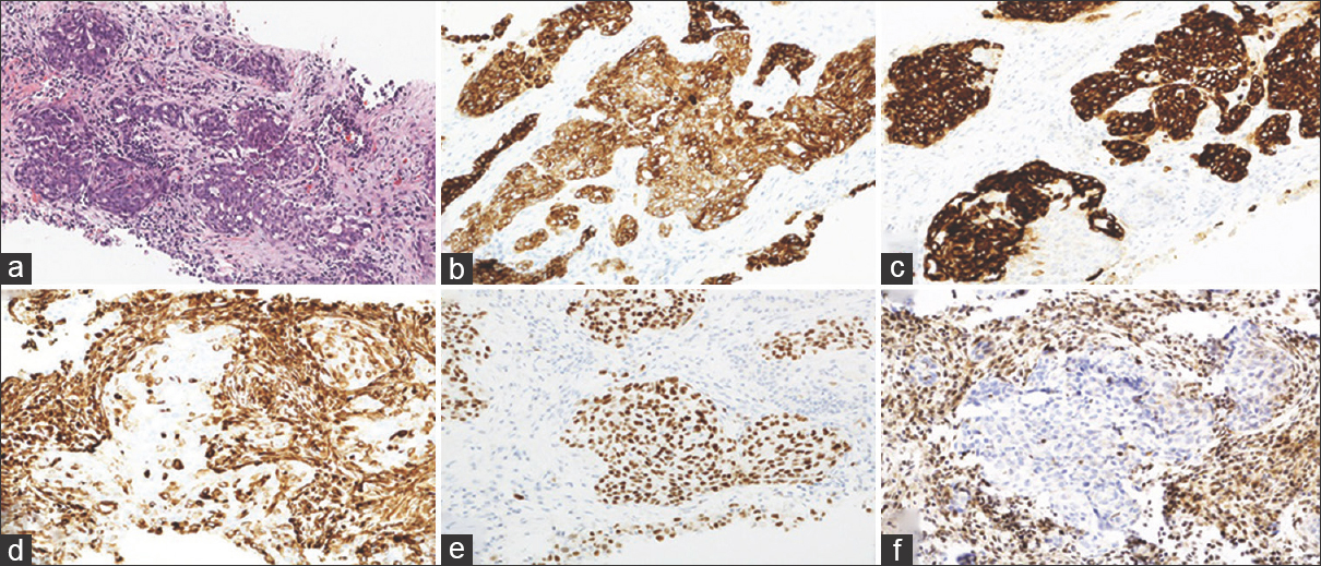
H and E and immunohistochemistry (IHC) results of the core biopsy in our case are presented in Figure 2a–f. In addition, TTF-1 and Napsin-A IHC performed on the core biopsy were negative.

- (a) Nests of malignant cells in a background of desmoplastic stroma and acute and chronic inflammation (H and E, ×200). (b) CK7 demonstrates diffuse cytoplasmic staining (×400). (c) CK20 demonstrates strong cytoplasmic staining (×400). (d) Vimentin demonstrates patchy positive staining in malignant cells (×400). (e) PAX8 demonstrates diffuse nuclear staining (×400). (f) INI-1 demonstrates loss of nuclear staining (×400)
ADDITIONAL QUIZ QUESTIONS
Q1. Which of the following statements is FALSE about renal medullary carcinoma?
-
It affects predominantly young patients of African descent
-
It is an aggressive tumor with a poor prognosis
-
It arises predominantly in the right kidney
-
High partial oxygen pressure and alkaline pH of the renal medulla in patients with sickle cell trait contribute to the mutagenesis and tumor formation.
Q2. RMC is thought to arise from the:
-
Proximal renal tubules
-
Distal renal tubules
-
Distal/terminal collecting ducts
-
Urothelium.
Q3. Which of the following IHC panels is characteristic of RMC?
-
HMWCK−, INI-1−, Vimentin+, PAX8+
-
HMWCK+, INI-1−, Vimentin+, PAX8−
-
HMWCK−, INI-1+, Vimentin+, PAX8+
-
HMWCK+, INI-1+, Vimentin−, PAX8+
Q4. What other tumors typically demonstrate INI-1 staining pattern that is similar to RMC?
-
Extrarenal malignant rhabdoid tumor
-
Atypical teratoid/rhabdoid tumor
-
Epithelioid sarcoma
-
All of the above.
ANSWERS TO ADDITIONAL QUIZ QUESTIONS
Q1 (d); Q2 (c); Q3 (a); Q4 (d)
Q1 (d): RMC is an aggressive malignant tumor with a poor prognosis. It is seen predominantly in young patients of African descent with sickle cell trait. Greater than 75% of RMC originate in the right kidney.[12] The low partial oxygen pressure and acid pH of the renal medulla promote chronic hemoglobin sickling and contribute to chronic ischemia, hypoxia, and vaso-occlusion, which are thought to promote mutagenesis.[10]
Q2 (c): RMC is thought to arise from the epithelium of the distal/terminal collecting ducts.
Q3 (a): RMC is positive for cytokeratin AE1/AE3, low molecular weight cytokeratin, vimentin, and PAX8. It is negative for HMWCK and demonstrates loss of nuclear INI-1 (hSNF5/SMARCB1/BAF47) staining. Staining for CK7 and CK20 is variable ranging from no staining to diffuse staining.
Q4 (d): INI-1 is a highly conserved factor in ATP-dependent chromatin remodeling complex. In addition to RMC, other tumors with absent INI-1 are pediatric renal and extrarenal malignant rhabdoid tumors, atypical teratoid/rhabdoid tumors of the central nervous system, and epithelioid sarcomas. Some epithelioid MPNST, a proportion of myoepithelial carcinoma, and EMCS also show loss of INI-1 staining.[11]
BRIEF REVIEW OF THE TOPIC
RMC arises in the renal medulla. It was first described in 1995 by Davis et al.,[14] who dubbed it as the “seventh sickle cell nephropathy;” the other six being hematuria, papillary necrosis, nephrotic syndrome, renal infarction, inability to concentrate urine, and pyelonephritis. These latter six pathologic changes seen in the renal medulla are associated with the chronic sickling process. The acid pH and hyperosmolar microenvironment of the medulla contribute to the increase in intracellular concentration and polymerization of hemoglobin S. The partial oxygen pressure in the renal medulla is 35–40 mmHg, below the 45 mmHg threshold for sickling. All these factors are thought to promote sickling not only in individuals with sickle cell disease but also in those with sickle cell trait. The pathologic sequelae of chronic sickling include chronic ischemia, hypoxia, and vaso-occlusion, all of which may promote mutagenesis and neoplastic transformation in the damaged renal medulla.[10]
RMC is thought to arise from the epithelium of the distal/terminal collecting ducts and has been proposed as a variant of CDC of the kidney. It has been hypothesized that chronic sickling increases levels of hypoxia-inducible factor (HIF), a transcription factor that regulates the expression of various genes. It induces TP53, which regulates cell death through apoptosis. In tumors lacking TP53, HIF induces vascular endothelial growth factor, promoting neovascularization, thus causing cancer progression.[10] Yang et al. studied molecular profiling of RMC using comparative genomic hybridization comparing the expression pattern of different genes in renal medullary carcinoma with all other types of renal tumors. They discovered that renal medullary carcinoma clustered most closely with urothelial carcinoma. Both have markedly elevated extracellular matrix genes, such as laminin alpha 3 and gamma 2, fibronectin 1, collagen Type III, and fibulin 2. Several genes are overexpressed in RMC, such as topoisomerase II, macrophage-stimulating 1 receptor, and angiogenesis-related genes, including peroxisome proliferator-activated receptor gamma angiopoietin -related gene.[15]
The common presenting complaints are hematuria and flank pain; whereas respiratory issues from bulky disease or pleural effusion are typically seen in <10% of presentations.[2] Interestingly, despite the left kidney being the most common location of benign hematuria, >75% of RMC originate in the right kidney.[12] Metastases at presentation are frequent. The most common metastatic sites include lymph nodes, lungs, liver, adrenal glands, and bone.[1] Some patients first present with signs of metastatic disease as was seen in our patient who presented with cough and shortness of breath.
RMC shows a variable architectural pattern including cribriform, reticular, yolk sac, adenoid cystic-like, trabecular, infiltrating solid cords, or papillae with central fibrovascular cores. Stroma is hypocellular, myxoid, edematous, with desmoplastic reaction and inflammatory infiltrate.[161718] Cytologically, the tumor cells are arranged in loosely cohesive clusters or dispersed singly. They have pleomorphic nuclei, prominent nucleoli, irregular nuclear membranes, distinct cell borders, and cytoplasmic vacuoles and often demonstrate rhabdoid features. Inflammation, stromal fragments, and sickle red blood cells may be present in the background.[345678919] The tumor cells are positive for mucin, typically positive for CK7, and vimentin (focal) and are negative for HMWCK, with only a few cases reported as positive for ulex europaeus agglutinin 1 lectin (UEA-1).
Morphologically, RMC can be confused with CDC as both tumors arise in the renal medulla. In fact, some publications described RMC as a subtype of CDC. Clinical presentation, morphology, and immunostains help differentiate these two tumor types. CDC occurs in older patients and is not associated with sickle cell trait/disease. It demonstrates tubular, tubulopapillary, and glandular structures and solid and/or nested growth patterns. Intraluminal basophilic to amphophilic mucin may be present. Stroma is myxoid to sclerotic with desmoplastic stromal reaction with or without inflammation.[16] Cytologic preparations are variably cellular and demonstrate medium-sized tumor cells with high nucleocytoplasmic ratios, large pleomorphic nuclei, coarse chromatin, inconspicuous to prominent nucleoli, and scant to moderate finely granular cytoplasm. Occasionally, intracytoplasmic mucin may be present.[19] The tumor cells are positive for HMWCK, UEA-1,[1020] and generally CK7, but not CK20.
High-grade urothelial carcinoma and RMC have some morphologic similarities, most notably the infiltrating growth pattern. The cells have relatively dense cytoplasm and nuclei with obvious malignant features, including pleomorphism, course chromatin, and prominent nucleoli.[19] Urothelial carcinoma is positive for HMWCK and p63, and many tumors are positive for both CK7 and CK20. RMC shows variable staining with CK7 and CK20 and is HMWCK and p63 negative.
The differential diagnosis of RMC also includes high-grade conventional clear cell RCC, which can grow in a variety of patterns including solid, alveolar, and acinar with numerous thin-walled blood vessels. Cytologically, high-grade RCC is arranged in clusters or sheets, sometimes with floral groups or short papillae that occasionally contain metachromatic basement membrane-like material. The cells have delicate wispy or finely vacuolated cytoplasm and centrally or eccentrically located large, pleomorphic nuclei with prominent nucleoli.[19] Like RMC, high-grade RCC may show rhabdoid features; however, it is typically negative for both CK7 and CK20. In addition, the majority of RMC show loss of nuclear staining for INI-1 whereas the staining is retained in RCC, UC, and CDC, even those with rhabdoid morphology.
Treatment for RMC is challenging due to the rarity of this tumor with a mean survival of less than a year. Neither chemotherapy nor radiation therapy is particularly effective. The current therapeutic approach with these aggressive tumors is radical nephrectomy followed by cytotoxic chemotherapy, but the prognosis remains dismal. A few newer treatment approaches including both cytotoxic chemotherapy and targeted therapy regimens have been shown to slightly increase average survival in small cohorts.[2021]
SUMMARY
Reported cases of RMC describing cytologic features and immunohistochemical characteristics are few and without long-term follow-up. A high index of suspicion in patients with known risk factors might prove to be helpful in early detection. Further studies with larger patient cohorts are needed to understand this rare malignancy.
COMPETING INTERESTS STATEMENT BY ALL AUTHORS
All authors declare that they have no competing interests.
AUTHORSHIP STATEMENT BY ALL AUTHORS
MM collected the details of the case, carried out literature review and drafted and edited the manuscript. MP collected the details of the case, performed photomicrographs, additional literature review and edited the manuscript. KM was involved in conceptualization and edited the manuscript. JT helped edit the manuscript. SH helped edit the manuscript. EL conceptualized the quiz case, performed additional literature review, and edited the manuscript. All authors read and approved the final manuscript.
ETHICS STATEMENT BY ALL AUTHORS
This report does not require approval from Institutional Review Board.
LIST OF ABBREVIATIONS (In alphabetic order)
CT − Computed tomography
EMCS − Extraskeletal myxoid chondrosarcoma
H and E − hematoxylin and eosin
HIF – Hypoxia inducible factor
HMWCK − High molecular weight cytokeratin
IHC – Immunohistochemistry
MPNST − Malignant peripheral nerve sheath tumor
RCC − Renal cell carcinoma
RMC – Renal medullary carcinoma
UEA 1 − Ulex europaeus agglutinin 1 lectin.
EDITORIAL/PEER-REVIEW STATEMENT
To ensure the integrity and highest quality of CytoJournal publications, the review process of this manuscript was conducted under a double-blind model (authors are blinded for reviewers and vice versa) through automatic online system.
REFERENCES
- Renal medullary carcinoma and sickle cell trait: A systematic review. Pediatr Blood Cancer. 2015;62:1694-9.
- [Google Scholar]
- Clinical outcome and prognostic factors in renal medullary carcinoma: A pooled analysis from 18 years of medical literature. Can Urol Assoc J. 2015;9:E172-7.
- [Google Scholar]
- Fine needle aspiration cytology diagnosis of renal medullary carcinoma: A case report. Acta Cytol. 2001;45:735-9.
- [Google Scholar]
- Cytology of metastatic renal medullary carcinoma in pleural effusion: A study of two cases. Diagn Cytopathol. 2009;37:843-8.
- [Google Scholar]
- Renal medullary carcinoma: A report of 2 cases and review of the literature. Arch Pathol Lab Med. 2003;127:e135-8.
- [Google Scholar]
- Pathologic quiz case: A 33-year-old man with an abdominal mass. Arch Pathol Lab Med. 2000;124:1561-3.
- [Google Scholar]
- Renal medullary carcinoma: Report of a case with positive urinary cytology. Diagn Cytopathol. 1998;18:276-9.
- [Google Scholar]
- Cytodiagnosis of renal medullary carcinoma: Report of a case with immunocytochemistry. Cytopathology. 2015;26:328-30.
- [Google Scholar]
- Renal medullary carcinoma: Clinical, pathologic, immunohistochemical, and genetic analysis with pathogenetic implications. Urology. 2002;60:1083-9.
- [Google Scholar]
- INI1-deficient tumors: Diagnostic features and molecular genetics. Am J Surg Pathol. 2011;35:e47-63.
- [Google Scholar]
- Renal medullary carcinoma: A case report and brief review of the literature. Ochsner J. 2014;14:270-5.
- [Google Scholar]
- Renal medullary carcinoma: Ultrastructural studies may benefit diagnosis. Ultrastruct Pathol. 2008;32:252-6.
- [Google Scholar]
- Renal medullary carcinoma. The seventh sickle cell nephropathy. Am J Surg Pathol. 1995;19:1-11.
- [Google Scholar]
- Gene expression profiling of renal medullary carcinoma: Potential clinical relevance. Cancer. 2004;100:976-85.
- [Google Scholar]
- Carcinoma of the collecting ducts of Bellini and renal medullary carcinoma: Clinicopathologic analysis of 52 cases of rare aggressive subtypes of renal cell carcinoma with a focus on their interrelationship. Am J Surg Pathol. 2012;36:1265-78.
- [Google Scholar]
- Renal medullary carcinoma: Report of seven cases from Brazil. Mod Pathol. 2007;20:914-20.
- [Google Scholar]
- Renal medullary carcinoma: Molecular, immunohistochemistry, and morphologic correlation. Am J Surg Pathol. 2013;37:368-74.
- [Google Scholar]
- Kidney. In: Demay RM, ed. The Art & Science of Cytopathology (2nd ed). Chicago: American Society for Clinical Pathology Press; 2012.
- [Google Scholar]
- Management and outcomes of patients with renal medullary carcinoma: A multicentre collaborative study. BJU Int. 2017;120:782-92.
- [Google Scholar]
- Topoisomerase II alpha status in renal medullary carcinoma: Immuno-expression and gene copy alterations of a potential target of therapy. J Urol. 2009;182:735-40.
- [Google Scholar]


