


Translate this page into:
Cytomorphology of columnar cell variant of papillary carcinoma thyroid: A case report and review of the literature
*Corresponding author
-
Received: ,
Accepted: ,
This is an open-access article distributed under the terms of the Creative Commons Attribution-Noncommercial-Share Alike 3.0 Unported, which permits unrestricted use, distribution, and reproduction in any medium, provided the original work is properly cited.
This article was originally published by Medknow Publications & Media Pvt Ltd and was migrated to Scientific Scholar after the change of Publisher.
Abstract
A 58 years old lady reported with history of progressively increasing lump in the neck. Patient had earlier undergone sub-total thyroidectomy (details not available) in a private institute one year back. Fine needle aspiration cytology (FNAC) of the present lump revealed features of papillary carcinoma thyroid. Patient subsequently underwent total thyroidectomy along with excision of a tumor nodule in the larynx. Gross examination of the specimen revealed a tumor nodule in the right lobe of the thyroid. Microscopic examination of the tumor nodule in the thyroid and larynx revealed a columnar cell variant of papillary carcinoma thyroid. Very few reports describing the cytomorphologic features of this variant of papillary carcinoma are available in the published literature. These reports highlight the absence or paucity of nuclear grooves and intranuclear inclusions in this variant. We describe a case of columnar cell variant of papillary carcinoma where nuclear grooves were prominently seen. In addition, we report the occurrence of rosette-like structures which were brought out better on FNA smears. These rosette-like structures have not been emphasized earlier in the published literature. The cytomorphologic features of this rare variant are also reviewed in this report.
Keywords
Columnar cell variant
fine needle aspiration cytology
papillary carcinoma thyroid

INTRODUCTION
Columnar cell variant of papillary carcinoma thyroid is a very rare neoplasm. The entity was first described by Evans in 1986.[1] It is an aggressive tumor associated with widespread dissemination and a fatal outcome. The cytology of the neoplasm is quite elusive since the various classical nuclear features of papillary carcinoma like ground glass nuclei, nuclear grooves and intranuclear inclusions have not been found to be consistently present in the published reports on this neoplasm.[23456] Biological behavior and histomorphology of this neoplasm is quite dissimilar to the conventional papillary carcinoma thyroid and hence it demands attention. The neoplasm consists of papillary fronds and follicles lined by columnar cells with pseudostratification, hyperchromasia of the nuclei and clear or vacuolated cytoplasm.[789] This variant of papillary carcinoma usually shows aggressive behavior with invasion of the surrounding soft tissue and even distant metastasis.[8] Herein, we report the fine-needle aspiration (FNA) features of a case of columnar cell variant of papillary carcinoma thyroid, which had cytological features that have not been observed in the earlier published cases.
CASE REPORT
A 58-year-old female patient presented with a history of progressively increasing lump in the neck. Review of past medical documents revealed that she had undergone sub-total thyroidectomy 1 year back in a private institute. Slides and blocks were not available for review. FNA was performed on the present lump. Papanicolaou and May Grünwald Giemsa stained smears were available. A cytological diagnosis of papillary carcinoma of thyroid was rendered based upon architectural characteristics and nuclear features. Patient underwent total thyroidectomy. The tumor was found to invade the larynx. Following the histopathological diagnosis of columnar cell variant of papillary carcinoma thyroid, a retrospective review of cytomorphology was performed.
Cytopathologic findings
The smears were cellular and showed tumor cells in aggregates and present singly. The tumor cells were arranged in papillary fronds, syncytial sheets and at places in a rosette-like pattern. The papillary fronds, periphery of syncytial fragments as well as the rosette-like clusters of tumor cells showed prominent pseudostratification [Figure 1a] of the nuclei. Many singly lying cells were also seen having a columnar configuration and wispy cytoplasm [Figure 1b, inset]. There was evidence of nucleomegaly, moderate degree of anisonucleosis, stippled chromatin, and the presence of occasional inconspicuous nucleoli. However, nuclear grooves [Figure 1c] were frequently encountered in the tumor cells. Intranuclear cytoplasmic inclusions were conspicuous by their absence. Mitotic figures were seen [Figure 1c, inset]. Rosette like clusters of the tumor cells were also observed having peripherally aligned nuclei and columnar cytoplasmic processes merging toward the center [Figure 1b].
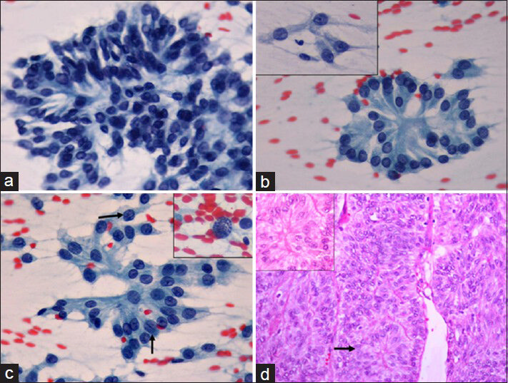
- (a) A papillary cluster of tumor cells displaying prominent pseudostratification of nuclei toward the periphery (Papanicolaou, ×400). (b) Rosette-like clusters of tumor cells with tall cytoplasm merging toward the lumen. Inset shows singly lying cells with columnar configuration and wispy cytoplasm (Papanicolaou, ×400). (c) Prominent nuclear grooves (black arrows) were seen in the fine needle aspiration smears. Inset shows presence of mitotic figures (Papanicolaou, ×400). (d) Histologic section of tumor showing trabecular arrangement with presence of pseudostratification and rosette-like structures (black arrow). Inset highlights the same rosette-like structures (H and E, ×200)
Histopathologic findings
The gross examination of the specimen revealed a thyroid gland 30 g in weight. Right lobe had a nodule 3.5 cm × 3.0 cm × 2.5 cm. The cut surface of the nodule in the thyroid was solid and homogenous. Microscopic examination revealed an encapsulated tumor mass with invasion of the capsule. There were areas of necrosis. The tumor cells had mostly trabecular arrangement with pseudostratification of nuclei [Figure 1d]. Cells were columnar and had clear to pale pink cytoplasm. Rosette-like structures were also noted in the broad trabeculae of columnar cells [Figure 1d, black arrow and inset]. The nuclei showed mild to moderate anisokaryosis with speckled nuclear chromatin. Single conspicuous nucleoli were visible in few cells with many nuclei showing presence of nuclear grooves. However, no intranuclear cytoplasmic inclusions or psammoma bodies were identified. The tumor showed frequent mitotic figures averaging 4-5/10 hpf. The nodule excised from the larynx showed a tumor mass with similar histomorphology. Immunohistochemistry performed showed diffuse positivity for thyroglobulin and carcinoembryonic antigen. The cells were negative for CK19, chromogranin, and synaptophysin. The case was diagnosed as columnar cell variant of papillary carcinoma thyroid.
DISCUSSION
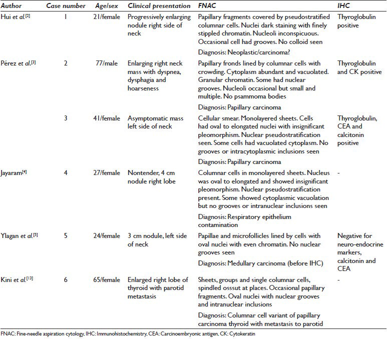
Fine-needle aspiration of the thyroid did not gain acceptance till early 1980s when Scandinavian investigators firmly reported its diagnostic accuracy.[10] Columnar cell variant of papillary carcinoma thyroid is one of the rarest morphological subtypes.[11] Earlier this subtype of papillary carcinoma was thought to be always aggressive; however, an encapsulated tumor appears to have a favorable outcome after complete excision.[12] There have been very few published reports on the cytomorphologic description of this rare variant of papillary carcinoma. Six case reports of FNA findings are available in the cytological literature.[234513] The details of these isolated cases have been highlighted in Table 1. The features of columnar cell variant of papillary carcinoma thyroid are elusive and hence the diagnosis is difficult solely based on FNA. All the five earlier published cases were diagnosed primarily on thyroid FNA, and the present case could not be diagnosed as a case of columnar cell variant of papillary carcinoma thyroid on FNA.[2345]
Pseudostratifcation of nuclei is a prominent feature of this neoplasm, and its presence should alert a cytopathologist regarding this variant of papillary carcinoma. Pseudostratification was present in all the published cases and also seen in our case. Tall cell variant of papillary carcinoma, which is the main differential diagnosis, lacks the feature of pseudostratification though it also has tall columnar cells. The tall cell variant has a single layer of columnar cells with deeply eosinophilic cytoplasm, which line the papillary fronds. The nuclear characteristics in tall cell variant are those of papillary carcinoma thyroid.[14] FNA findings in two cases of combined columnar and tall cell variant of papillary carcinoma have also been reported.[1415] Nuclear grooves were present frequently in our case and one case of mixed columnar cell and tall cell features in contrast to their absence in three of the earlier reported cases and their occasional presence in the other two published cases.[234515] Intranuclear cytoplasmic inclusions were absent in all the published cases except in two cases reported by Kini et al. and Tranchida et al.[23451315] Finely stippled chromatin and inconspicuous nucleoli in our case were in agreement with the finding in most of the published cases.[2345] The present case also had cells scattered singly and in small groups of 2-3 cells with columnar morphology and a wispy cytoplasm. Presence of this feature in the case of papillary carcinoma may also give a clue about the possibility of this variant. Our case in addition also displayed a rosette-like arrangement of columnar tumor cells. These structures were highlighted better in the FNA smears as compared to the histologic sections. To the best of our knowledge, the presence of these rosette-like structures formed by the merging of tall columnar cytoplasm, have not been emphasized earlier in histologic sections or FNA cytology (FNAC) of this tumor.
Our patient had recurrence within 1 year, and the recurrent tumor showed invasion of the capsule and extension into the larynx. Poorer prognosis and aggressive behavior of this variant of papillary carcinoma warrants an early recognition of the tumor. FNAC being the first line of investigation in the diagnosis of thyroid swellings, recognition of the cytologic characteristics of this aggressive subtype may be of utility in the management of these patients.
COMPETING INTERESTS
The authors declare that they have no competing interests.
AUTHORSHIP STATEMENT BY ALL AUTHORS
Each author acknowledges that this final version was read and approved. All authors qualify for authorship as defined by ICMJE http://www.icmje.org/#author. Each author participated sufficiently in the work and takes public responsibility for appropriate portions of the content of this article.
ETHICS STATEMENT BY ALL AUTHORS
As this is a case report without identifiers, our institution does not require approval from Review Ethics Board (REB).
EDITORIAL/PEER-REVIEW STATEMENT
To ensure the integrity and highest quality of CytoJournal publications, the review process of this manuscript was conducted under a double blind model (authors are blinded for reviewers and vice versa) through automatic online system.
REFERENCES
- Columnar-cell carcinoma of the thyroid. A report of two cases of an aggressive variant of thyroid carcinoma. Am J Clin Pathol. 1986;85:77-80.
- [Google Scholar]
- Columnar cell carcinoma of the thyroid. Fine needle aspiration findings in a case. Acta Cytol. 1990;34:355-8.
- [Google Scholar]
- Fine-needle aspiration cytology of columnar-cell carcinoma of the thyroid: Report of two cases with cytohistologic correlation. Diagn Cytopathol. 1998;18:352-6.
- [Google Scholar]
- Cytology of columnar-cell variant of papillary thyroid carcinoma. Diagn Cytopathol. 2000;22:227-9.
- [Google Scholar]
- Columnar cell variant of papillary thyroid carcinoma. Report of a case with cytologic findings. Acta Cytol. 2004;48:73-7.
- [Google Scholar]
- Columnar cell carcinoma of the thyroid gland. A case report and review of the literature. Hum Pathol. 1995;26:691-3.
- [Google Scholar]
- The columnar cell variant of thyroid papillary carcinoma. Case report and discussion of an unusually aggressive thyroid papillary carcinoma. Am J Surg Pathol. 1995;19:940-7.
- [Google Scholar]
- Thyroid papillary carcinoma of columnar cell type: A clinicopathologic study of 16 cases. Cancer. 1998;82:740-53.
- [Google Scholar]
- Fine-needle aspiration of follicular lesions of the thyroid. Diagnosis and follow-up. Cytojournal. 2006;3:9.
- [Google Scholar]
- Prognostic significance of histologic grading compared with subclassification of papillary thyroid carcinoma. Cancer. 2000;88:1902-8.
- [Google Scholar]
- Encapsulated columnar-cell neoplasms of the thyroid. A report of four cases suggesting a favorable prognosis. Am J Surg Pathol. 1996;20:1205-11.
- [Google Scholar]
- Solitary parotid metastasis from columnar cell carcinoma of the thyroid: A diagnostic dilemma. Diagn Cytopathol. 2003;28:72-5.
- [Google Scholar]
- Mixed columnar cell and tall cell variant of papillary carcinoma of thyroid: A case report and review of the literature. Pathology. 2000;32:286-9.
- [Google Scholar]
- Preoperative cytologic diagnosis of papillary thyroid carcinoma with mixed columnar cell and tall cell features. Diagn Cytopathol. 2012;40(Suppl 1):E4-7.
- [Google Scholar]


