Translate this page into:
Cytomorphological spectrum of epididymal nodules: An institution's experience
*Corresponding author
-
Received: ,
Accepted: ,
This is an open access article distributed under the terms of the Creative Commons Attribution-NonCommercial-ShareAlike 3.0 License, which allows others to remix, tweak, and build upon the work non-commercially, as long as the author is credited and the new creations are licensed under the identical terms.
This article was originally published by Medknow Publications & Media Pvt Ltd and was migrated to Scientific Scholar after the change of Publisher.
Abstract
Background:
Epididymal lesions are uncommon in clinical practice. Few case series has been described in the literature documenting the role of cytology in the evaluation of epididymal nodules. This study was undertaken to analyze the cytomorphology of epididymal nodules and to evaluate role of fine-needle aspiration biopsy (FNAB) in early definitive diagnosis of epididymal nodules.
Materials and Methods:
A total of seventy cases of epididymal nodules were aspirated over a period of 6 years in the Department of Pathology. These cases were taken from the cytology record as a part of this study. The aspiration was performed using 22/23-gauge needle. Smears were stained with May-Grunwald-Giemsa and Papanicolaou stains. Special stains and immunocytochemistry were performed, wherever required.
Results:
The cytological features were adequate to establish the diagnosis in sixty cases. The lesions diagnosed were tuberculosis 16 (22.85%), spermatoceles 12 (17.14%), benign cystic lesion 8 (11.42%), encysted hydrocele 8 (11.42%), acute suppurative lesion 6 (8.57%), filariasis 4 (5.71%), chronic epididymitis 3 (4.28%), nodular fasciitis 1 (1.42%), epidermal inclusion cyst 1 (1.42%), and cystic adenomatoid tumor 1 (1.42%). Ten cases were inadequate to establish the diagnosis. FNAB was useful in diagnosis of 86% of cases. Infectious lesions of the epididymis were more common than neoplastic lesions. Patients with infection responded well to medical treatment.
Conclusions:
FNAB is an easily available technique for palpable lesions of epididymis, and it helps in making an early, near definitive diagnosis of epididymal lesions. It also helps to avoid unnecessary surgical interventions and helps in timely management.
Keywords
Adenomatoid tumor
epididymis
tuberculosis
INTRODUCTION
The epididymis is a crescent-shaped organ intimately attached to the posterolateral aspect of the testis. It is a complex tubular network that connects the testicular efferent ducts to the vas deferens.[1] Variety of nonneoplastic and neoplastic lesions involves the epididymis.[2] Epididymis is the most commonly involved site for nonneoplastic extratesticular lesions of scrotum.[3] Most nonneoplastic lesions are managed conservatively. In such a setting, fine-needle aspiration biopsy (FNAB) plays an important diagnostic role so as to avoid unnecessary surgical interventions.[2]
The present retrospective study was done with the aim to study the cytomorphology of epididymal swellings and role of FNAB in early definitive diagnosis.
MATERIALS AND METHODS
A total of seventy cases of epididymal nodules aspirated over a period of 6 years from January 2010 to December 2015 in the Department of Pathology were studied. The aspiration was performed using 22/23-gauge needle and 10/20 ml syringe in supine position without local anesthesia. The smears were stained with May-Grunwald-Giemsa and Papanicolaou stains. Special stains and immunocytochemistry were performed, wherever required.
RESULTS
Seventy cases of epididymal nodules aspirated were in age range from 13 to 62. The most common age group was 20–30 followed by 31–40, 41–50, and 10–19. The most common clinical presentation was painless epididymal nodule followed by tender or painful epididymal nodule. Other complaints seen were fever, infertility, trauma, urinary complaints, and dragging sensation. FNAB was carried out in all cases, and adequate material was obtained. Diagnosis of all the cases was established on cytological features. Out of 70 cases, 58 cases were nonneoplastic, 10 were inadequate and 1 was reparative, and other was benign neoplastic lesion. The lesions diagnosed were tuberculosis 16 (22.85%), spermatoceles 12 (17.14%), benign cystic lesion 8 (11.42%), encysted hydrocele 8 (11.42%), acute suppurative lesion 6 (8.57%), filariasis 4 (5.71%), chronic epididymitis 3 (4.28%), nodular fasciitis 1 (1.42%), epidermal inclusion cyst 1 (1.42%), and cystic adenomatoid tumor 1 (1.42%). FNAB was useful in diagnosis of 86% of cases. No malignant lesion was observed in the present study.
Cytopathological findings
-
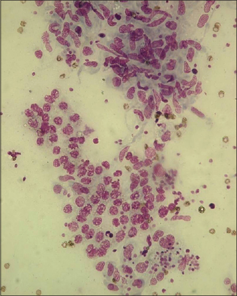
Tuberculous/granulomatous epididymitis (n = 16/70 cases): The patients with granulomatous epididymitis were in the age range of 15–35, and the most common age group was 20–30. The nodules were firm, nontender, indurated, and measured 2–3 cm in size. Smears prepared showed epithelioid cell granulomas with or without Langhans giant cells in 12 cases; caseous necrotic material and degenerating granulomas were seen in six cases [Figure 1]. Suppuration was present in three cases. Ziehl–Neelsen stain revealed acid-fast bacilli (AFB) in one case. All the cases were confirmed by culture and/or polymerase chain reaction
-
Acute suppurative lesion (n = 6/70): The patients were in age range of 10–40, and the most common age group was 20–30. These patients had firm swellings which were tender on palpation and measured 2–3 cm in size. The smear showed cluster of epithelial cells with polymorphs. All the cases were AFB negative. The suppuration was resolved after a course of antibiotics
-
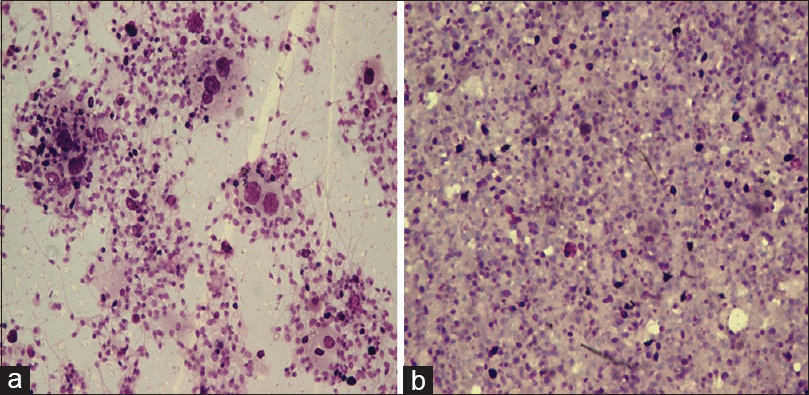
Spermatoceles (n = 12/70 cases): The patients were in age range of 20–45, and the most common age group was 20–30. These patients had cystic swellings which were nontender, 1–2 cm in size, and yielded clear straw-colored fluid. Smears prepared showed numerous sperms on a clear background. Few cystic macrophages were also seen, but inflammation was scant or absent. Lack of inflammation and foreign-body giant cells helped to distinguish the condition from a sperm granuloma [Figure 2a and b]
-
Benign cystic lesion (n = 8/70): The patients were in age range of 25–65, and the most common age group was 20–30. These patients had cystic swellings which were soft, nontender, 1–2 cm in size, and yielded clear straw-colored fluid. Smears prepared showed only cystic macrophages and epithelial cells
-
Encysted hydrocele (n = 8/70): The patients were in age range of 15–50, and the most common age group was 30–40. These patients had cystic swellings which were soft and nontender, 6 cm in size, and yielded clear straw- or amber-colored fluid [Figure 3]. Smears prepared showed few lymphocytes, macrophages, and mesothelial cells
-
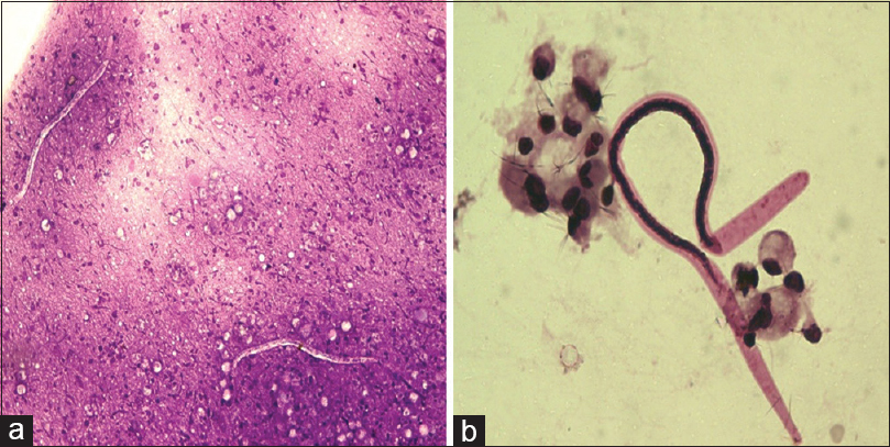
Filariasis (n = 4/70): The patients with filarial epididymitis were in age range of 20–30. The nodules were firm, nontender, indurated, and measured 2–3 cm in size. Smears prepared showed microfilarial worms in all the cases, and the background showed occasional histiocytes in one case and acute inflammatory background in other case [Figure 4a and b]
-
Chronic epididymitis (n = 3/70): The patients were in the age range of 20–35, and the most common age group was 20–30. These patients had swelling which was firm, nontender, and measured <1 cm in size. The smears revealed columnar epithelium with lymphocytes
-
Nodular fasciitis (n = 1/70): The patient was 40 years old with firm and nontender epididymal swelling 3 cm in size, and smear showed fibroblasts with ovoid-shaped nuclei, pale bland nuclear chromatin, and inflammatory cells
-
Epidermal inclusion cyst (n = 1/70): The patient was 40 years old with cystic epididymal swelling measuring 1 cm × 1 cm in size, and smear showed singly scattered and cluster of anucleate squames. Features were consistent with epidermal inclusion cyst
-
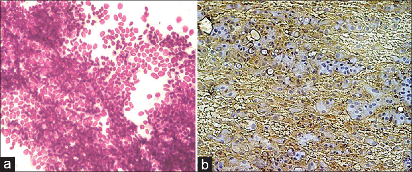
Cystic adenomatoid tumor (n = 1/70): The patient was 35 years old and presented with cystic swelling of epididymis which was firm in consistency and measured 5 cm × 4 cm in size, nontender on palpation. The cytospin and direct smears were cellular with the presence of monolayer sheets and cluster of small monotonous cells, the cells were round to oval with vesicular nuclei, with evenly distributed chromatin and small inconspicuous nucleoli [Figure 5a]. Few of cells were showing signet ring-like morphology. Tumor cells showed immunoreactivity with calretinin [Figure 5b]. The swelling was excised, and histopathological examination confirmed the diagnosis of cystic adenomatoid tumor.

- Epithelioid cell granulomas, sheet of epithelial cells, and spermatids (MGG, ×400)

- (a) Few cystic macrophages engulfing spermatozoa, numerous spermatids in a clear background (MGG, ×400). (b) Numerous spermatozoa, spermatids in a proteinaceous background with no inflammation (MGG, ×400)

- Encysted swelling filled with serous fluid

- (a) Microfilaria in an acute inflammatory background (MGG, ×400). (b) Microfilaria with background showing few histiocytes (MGG, ×400)

- (a) The cytospin smears were cellular with the presence of monolayer sheets; cells were round to oval with vesicular nuclei, small inconspicuous nucleoli along with stromal fragments (MGG, ×400). (b) Tumor cells showed immunoreactivity with calretinin (IHC, ×400)
DISCUSSION
Epididymal nodules are not infrequently encountered in surgical practice. FNAB of lesions is not easy due to their smaller size and slippery nature; however, it is the preferred method to evaluate the epididymal nodular swelling because it is rapidly done and less traumatic than a biopsy.[4]
FNAB of epididymal nodules plays an important role to differentiate nonneoplastic lesions from neoplastic lesions since most of the nonneoplastic lesions are conservatively managed. FNAB also avoids unnecessary surgical intervention and influences the management strategies.[5]
Various inflammatory, benign, and malignant lesions of epididymis have been described in the literature. The tubercular lymphadenitis is the most common form of extrapulmonary tuberculosis followed by genitourinary tuberculosis,[6] comprising 30% cases of nonpulmonary tuberculosis.[7] The epididymis is one of the most commonly involved sites for genital tuberculosis in males.[8] The epididymal tuberculosis accounts for about 20% of genitourinary tuberculosis.[9] The epididymal tuberculosis has been reported in young adults, with median age of 32 years and age range of 21–37 years in a review of 40 cases of isolated epididymal tuberculosis by Viswaroop et al.[10] The isolated epididymal tuberculosis occurs unilaterally, but bilateral involvement has also been reported by Viswaroop et al.[10] In the present study, the most common age group was 20–30 years with unilateral presentation in 13 out of 16 cases. Tuberculous epididymitis results from hematogenous spread with predominant caudal involvement attributed to high vascularity.[11] Higher frequency of isolated lesions in children favored the possibility of hematological spread of infection, while in adult, tuberculous epididymo-orchitis occurs as a result of direct spread from the urinary tract.[12] FNAB serves as a minimally invasive technique in the diagnosis of tuberculous epididymitis and provides adequate material for cytological and microbiological examination, avoiding unnecessary surgeries.[13] The cytological diagnosis of tuberculous epididymitis prompts a search for genitourinary tuberculosis.[14] The differential diagnosis includes spermatic granuloma and spermatoceles. The spermatic granuloma smears will show epithelioid granulomas and macrophages engulfing spermatozoids, tailless spermatozoids, and inflammatory cells. Caseous necrosis is not seen.[15] The aspirate is milky in spermatoceles, with the presence of numerous immobile spermatozoids, histiocytes, and spermiophage. A spermatocele can often be completely evacuated by fine-needle aspiration, leaving no residual lesion.[2]
Microfilarial infection is the second common infection encountered in epididymis in an Indian scenario.[16] The adult worms usually reside in lymphatic vessels of the epididymis, spermatic cord, paratesticular region, and juxtatesticular lymphatic vessels in “nests” preferential site.[17] The present study showed both coiled and uncoiled microfilariae in a clean and acute suppurative background on smear from epididymal nodules. Possibility of parasite should be ruled out in the presence of eosinophils in aspirate smear from epididymal nodule. Acute and chronic epididymo-orchitis smears reveal acute, chronic inflammatory response without demonstrable foreign bodies, infectious agents, or granuloma formation.
Hydrocele presents as a concentric swelling with pale or dark amber aspirate. A pale aspirate is sparsely cellular with few mesothelial cells and lymphocytes. A dark amber sample contains typical and reactive mesothelial cells singly or in clusters, histiocytes, and debris, representing a hydrocele of long period.[2] Fine-needle aspiration is both diagnostic and therapeutic in hydroceles and spermatoceles. In the present study, pale-colored aspirate with smear showing occasional mesothelial cells and lymphocytes was seen.
Hematocele is considered when altered blood is aspirated with antecedent history of trauma.[4]
Adenomatoid tumor is the most common benign tumor of paratesticular tissue and accounts for 32% of all tumors in epididymis. The structural and immunohistochemical studies support the mesothelial origin.[18]
On FNAB, adenomatoid tumors reveal abundant cellularity with sheets and multilayered clusters of monotonous cells with round or ovoid, eccentric nuclei containing small central nucleoli. The paranuclear clearing with a pink coloration on Giemsa stain or a clear vacuole-like area on Papanicolaou stain with a background of naked nuclei and stromal fragments are seen.[19] The cytological differential diagnosis includes reactive mesothelial hyperplasia; however, the usual coexistence of chronic inflammatory cells and fibrosis suggests hyperplasia. The cells show anisocytosis, pleomorphism, nuclear grooves, lobulation, amitosis, and moderate amount of pale cytoplasm can be seen. Spermatic granuloma shows histiocytes filled with sperm heads and debris in the background. Cytologically papillary cystadenoma comprises papillary structures and single isolated cells without nuclear atypia and with cytoplasmic vacuoles in a mucoid background. Malignant mesothelioma shows a diffuse cell pattern with cells having ruffled borders with dense cytoplasm, nuclear enlargement, macronucleation, and multinucleation. Metastatic adenocarcinoma shows cytological features of malignancy, and cells are positive for mucicarmine.[2021] In the present study, smears revealed epithelial-like cells in sheets and discohesive clusters of monotonous cells with ovoid eccentric nuclei containing small central nucleoli with occasional scattered spindle cells. Tumor cells showed immunoreactivity with calretinin. The swelling was excised, and histopathological examination showed features consistent with cystic adenomatoid tumor.
Primary lymphoma of epididymis is rare.[22] Few case reports of cytologic diagnosis of primary epididymal lymphoma have been described. Diagnosis of primary high-grade lymphomas is usually straightforward as they have overt cytological atypia.[22]
CONCLUSIONS
FNAB is an easily available technique for the palpable lesions of epididymis and helps in making an early, near definitive diagnosis of epididymal lesions. The cytological diagnosis of epididymal tuberculosis prompts a search for genitourinary tuberculosis. It also helps avert unnecessary surgical interventions and promotes timely management.
COMPETING INTERESTS’ STATEMENT BY ALL AUTHORS
The authors declare that they have no competing interests.
AUTHORSHIP STATEMENT BY ALL AUTHORS
All authors of this article declare that we qualify for authorship as defined by ICMJE http://www.icmje.org/#author. Each author has participated sufficiently in the work and takes public responsibility for appropriate portions of the content of this article. JNB carried out the procedure, interpreted the results, and drafted the manuscript. BD interpreted the results and helped to draft the manuscript. JB helped to carry out the procedure, collected the details of the case, and helped to draft the manuscript. SJ participated in the interpretation of the results and participated in its design and coordination. All authors read and approved the final manuscript. Each author acknowledges that this final version was read and approved.
ETHICS STATEMENT BY ALL AUTHORS
As this is a case report without identifiers, our institution does not require approval from Institutional Review Board (or its equivalent).
LIST OF ABBREVIATIONS (In alphabetic order)
AFB - Acid-fast bacilli
FNAB - Fine needle aspiration biopsy.
EDITORIAL/PEER-REVIEW STATEMENT
To ensure the integrity and highest quality of CytoJournal publications, the review process of this manuscript was conducted under a double-blind model. (authors are blinded for reviewers and vice versa) through automatic online system.
REFERENCES
- Epididymectomy. In: Graham SD, Keane TE, eds. Glenn's Urologic Surgery (7th ed). Philadelphia, USA: Lippincott Williams and Wilkins; 2010. p. :410.
- [Google Scholar]
- Aspiration cytology of palpable lesions of the scrotal content. Diagn Cytopathol. 1990;6:169-77.
- [Google Scholar]
- Clinico-radiological and pathological evaluation of extra testicular scrotal lesions. J Cytol. 2013;30:27-32.
- [Google Scholar]
- Fine needle aspiration of epididymal nodules in Chandigarh, North India: An audit of 228 cases. Cytopathology. 2006;17:195-8.
- [Google Scholar]
- Clinical presentation and diagnostic approach in cases of genitourinary tuberculosis. Indian J Urol. 2008;24:401-5.
- [Google Scholar]
- Incidence, etiopathogenesis and pathological aspects of genitourinary tuberculosis in India: A journey revisited. Indian J Urol. 2008;24:356-61.
- [Google Scholar]
- Genitourinary tuberculosis in pediatric surgical practice. J Pediatr Surg. 1997;32:1283-6.
- [Google Scholar]
- Isolated tuberculous epididymitis: A review of forty cases. J Postgrad Med. 2005;51:109-11.
- [Google Scholar]
- Tuberculous epididymitis as a cause of testicular pseudomalignancy in two young children. Pediatr Infect Dis. 1985;4:59-62.
- [Google Scholar]
- Fine needle aspiration cytology of tubercular epididymitis and epididymo-orchitis. Acta Cytol. 2006;50:243-9.
- [Google Scholar]
- Fine needle aspiration cytology versus open biopsy for evaluation of chronic epididymal lesions: A prospective study. Scand J Urol Nephrol. 2005;39:219-21.
- [Google Scholar]
- Spermatic granuloma. Diagnosis by fine needle aspiration cytology. Acta Cytol. 1989;33:1-5.
- [Google Scholar]
- The preferential site of adult Wuchereria bancrofti: An ultrasound study of male asymptomatic microfilaria carriers in Pondicherry, India. Natl Med J India. 2004;17:195-6.
- [Google Scholar]
- Aspiration cytology of adenomatoid tumor of epididymis: An important diagnostic tool. J Surg Case Rep. 2012;2012:11.
- [Google Scholar]
- Paratesticular adenomatoid tumors. The cytologic presentation in fine needle aspiration biopsies. Acta Cytol. 1989;33:6-10.
- [Google Scholar]
- Fine needle aspiration cytology of adenomatoid tumour – A case report with review of literature. Indian J Pathol Microbiol. 2005;48:503-4.
- [Google Scholar]


