


Translate this page into:
Virtual microscopy in cytotechnology education: Application of knowledge from virtual to glass
*Corresponding author
-
Received: ,
Accepted: ,
This is an open-access article distributed under the terms of the Creative Commons Attribution-Noncommercial-Share Alike 3.0 Unported, which permits unrestricted use, distribution, and reproduction in any medium, provided the original work is properly cited.
This article was originally published by Medknow Publications & Media Pvt Ltd and was migrated to Scientific Scholar after the change of Publisher.
Abstract
Background:
Virtual microscopy (VM) is a technology in which the glass slides are converted into digital images. The main objective of this study is to determine if cellular morphology, learned through virtual microscopy, can be applied to glass slide screening.
Materials and Methods:
A total of 142 glass slides (61 teaching and 81 practice) of breast, thyroid, and lymph node fine needle aspiration body sites were scanned with a single focal plane (at 40X) using iScanCoreo Au (Ventana, Tuscan, AZ, USA, formerly known as BioImagene, California, USA). Six students including one distant student used these digital images to learn cellular morphology and conduct daily screening. Subsequently, all the students were tested on 10 glass slides using light microscopy (LM). At the end of the study, the students were asked to respond to an online survey on their virtual microscopy experience. The glass slide screening test scores of the participating students who were taught through VM and tested on glass slides (VMLM group) were compared with the last three classes of students who were taught through LM and tested on glass slides (LMLM group).
Results:
A non-parametric statistical analysis indicated no difference (P = 0.20) in the glass screening test scores between VMLM (median = 93.5) and LMLM groups (median = 87). The survey indicated that the annotated teaching slides and access to the VM, off campus, were well appreciated by the students.
Conclusions:
Although the students preferred LM, they were able to apply the cytological criteria learned through VM to glass slide screening. Overall, VM was considered a great teaching tool.
Keywords
Cytotechnology
education
virtual microscopy

BACKGROUND
Virtual microscopy (VM) is a technology in which glass slides are scanned and converted into digital images.[1–3] These digital images are stored, viewed, and screened on the computer using a mouse. This technology is widely used in medical student education in pathology, continuing education, histopathology, cytology, veterinary and comparative pathology, and hematology.[4] To our knowledge, no one has studied if students are able to learn cytomorphology with VM and apply the knowledge to glass slide screening and interpretation using LM, particularly in cytology education, which is the main reason for conducting our study.
The cytotechnology program of the University of Nebraska Medical Center (UNMC) offers a distance learning option to students at our satellite location at the Carle Foundation Hospital in Urbana, IL. These students follow the same curriculum as the UNMC students; however, they have separate microscope teaching sessions, using different sets of glass slides for screening and interpretation, instruction, and practice. Of late, we have received many requests for either a part-time program or to study via distance from other states. These potential students may not have access to a cytology laboratory that can provide microscopic instruction. Considering these requests, we have decided to implement VM into our program, to improve the quality of education for students learning cytology, to standardize training / teaching on-campus, as well as for distance-learning students, to expand distance learning by creating online training programs, and to keep pace with the technological advances that will engage and challenge students.
Programs that utilize glass slides for teaching face well-documented challenges, which include breakage, loss of slides, and frustration on the part of the students for having to share glass slides among themselves.[5] In order to overcome such issues, many fields in which LM is in practice have already started implementing VM, and the benefits have been discussed in prior studies.[5–8] In most of the previous studies conducted in histology and medical education, the students’ responses to VM have been collected by means of a survey. Most of the student responses indicate that VM has enabled their learning, as it is an excellent resource, time efficient, easy to use, more productive for the students, more stimulating, flexible for learning, it eliminates the need to share glass slides with other students, and provides effective use of time.[9–13] To date, digital images have been used for telecytology,[14–25] training and education,[26] and proficiency testing.[27–29] In contrast to many of the educational programs in histology that have accepted VM,[26] the education programs in cytology have not yet completely embraced this technology.
This study was conducted to investigate whether VM could be utilized as the primary teaching tool for the basic cytomorphological criteria as well as screening and interpretation. The study also assessed the experiences of the UNMC Cytotechnology Program students in using VM. It was expected that the VM learning experience would allow the students to view images anywhere at any time, as many times as they wished, thus enhancing their learning experience. This was in contrast to the traditional microscope teaching sessions given by a faculty member in which students were given only a single viewing, as the sessions were not recorded and could not be replayed.
The overall objective of this study was to determine if learning through VM could be applied effectively to glass slide screening using LM, and to evaluate students’ opinions regarding the use of virtual slides compared to glass slides.
MATERIALS AND METHODS
Subjects of this study were students enrolled in the cytotechnology program at the UNMC and the distant / satellite program at the Carle Foundation Hospital in Urbana, Illinois. The students were sent an e-mail notification, briefly describing the study, and were given the choice to either volunteer for this study or not to participate. They were told that their scores of this study’s final glass slide screening test would not be included in their course grade. They were also informed that the entire set of glass slides that were digitized for this study would be taught using the multi-headed microscope in the traditional manner once the study was completed, and the data were collected if they felt it necessary. All students (n = 6) at UNMC and the Carle Foundation Hospital agreed to participate in the study and a signed informed consent was taken from each student, which was approved by the Institutional Review Board of the UNMC.
Slide selection
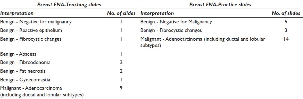
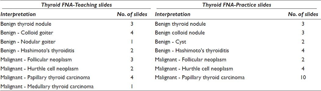
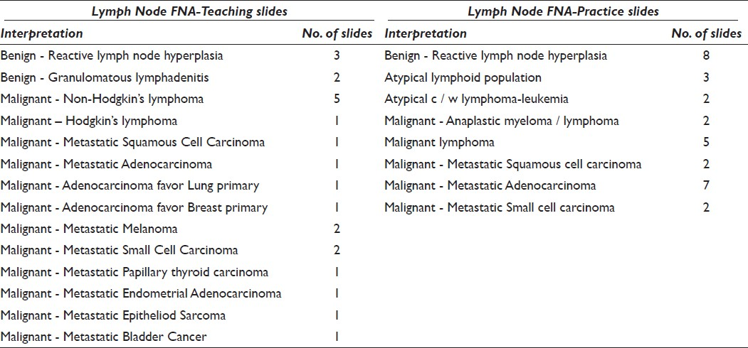
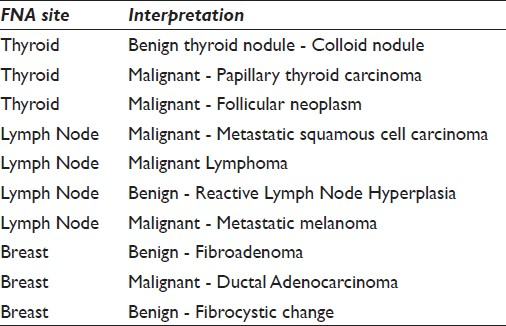
The cytotechnology program of UNMC has an extensive and diverse bank of gynecological and non-gynecological glass slides. These include both teaching and practice (unknown cases) glass slides. Teaching slides have cells already identified with dots and have the diagnosis and patient information written on a card that stays with the slide. The virtual teaching images have annotations, with cells already identified, cytomorphological criteria written next to the cells, patient information pertinent to the diagnosis, and references from the text books. The practice slides are glass slides that we assign to the students, to screen as unknowns. These slides have no dot-identifying cells and have no diagnosis or other information given, except the site. The virtual images of these slides do not have any annotations or other information given. The students must practice screening, identifying, and interpreting the cells of interest on the unknown images. For the purpose of this study, we selected 142 glass slides (61 teaching and 81 practice slides) from breast, thyroid, and lymph node fine needle aspiration (FNA) study sets [Tables 1–3]. Both teaching and practice slides were selected from all possible diagnostic categories found in the cytotechnology program files, ranging from benign to malignant. These slides are the same slides that had been used for teaching and practice in previous years.
The type of the slides included Papanicolaou-stained (a) membrane filter preparations, (b) cytocentrifuge preparations, and (c) aspirate smear slide preparations; Diff-Quik®-stained (a) cytocentrifuge preparations and (b) aspirate smear slide preparations; and Hematoxylin and Eosin-stained cell block preparation slides.
Given that whole slide digitization would take a longer time (scanning and screening) and required an extremely large storage file size,[30] only selected segments of the slides were scanned. All slides were pre-screened and a representative area that contained sufficient cells to provide an interpretation was marked. This representative area was not limited only to diagnostic cells; the students were still required to locate the abnormal / diagnostic cells of interest within a group of normal cells, in order to render an interpretation.
Digital image acquisition
Once the slides were de-identified and new labels were given, iScanCoreo Au (Ventana, Tucson, AZ, USA formerly known as Bioimagene, California, USA) was used to digitize the slides using a single plane (2D) at 40X magnification. The slides were loaded on the slide holder, and a thumbnail of the slide was produced. For each slide, the software of the instrument outlined the area of the sample to be scanned and chose the focal points. Once the focal points were chosen, the instrument scanned the slide using normal focus algorithms. The output file was saved in JPEG 2000 format, in a password protected, encrypted 2-Terabyte-capacity external hard drive (Seagate FreeAgent GoFlex Desk (2TB)). The average file size of the glass slides scanned was 426.523581 MB.
All digitized images were uploaded to the http://unmc.pathxchange.org website under a separate folder named Cytotechnology. The teaching virtual images were annotated extensively, emphasizing the important morphological criteria, diagnoses, clinical data, differential diagnosis, and references within the students’ textbooks. The cells of interest were highlighted by being boxed with a specific color and details about the cells of interest were entered into a text box that appeared on the left of the screen [Figure 1].
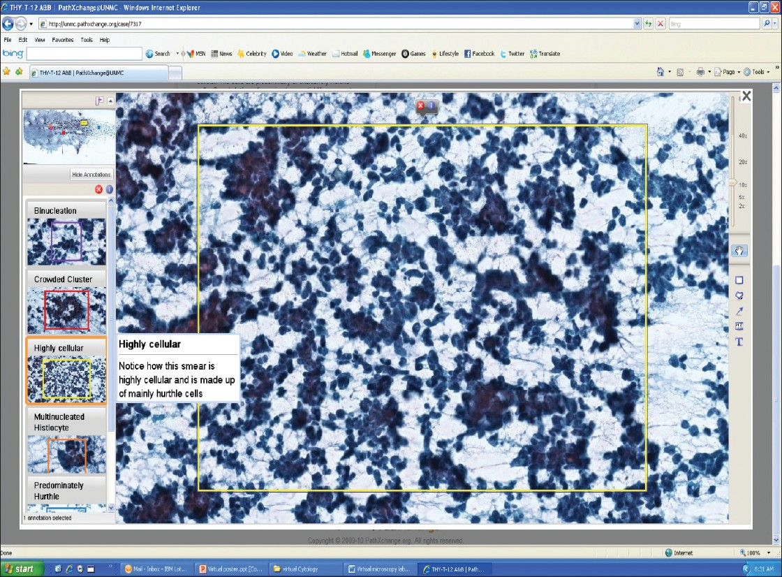
- This image is an example of an annotated teaching slide seen on the UNMC PathXchange website
At the beginning of the study, the students were given a detailed demonstration on the PathXchange website, images, annotation details, and screening procedures of the images, using various magnifications, and so forth. All students were given a username and password to log on to the website.
After the traditional lecture on breast FNA, the students were asked to view the teaching virtual images of the benign and malignant categories on the PathXchange website. Although the teaching slides were annotated and could go directly to the areas of interest, the students were encouraged to screen the entire image. Students also had options of screening the images with a range of magnification (2×, 5×, 10×, 20×, and 40×) by using a bar on the right side of the main image. The students were encouraged to discuss the annotations with each other while viewing the teaching images. There was no interaction with the faculty members, except during the traditional lectures for the course of this study. The students were instructed to rely only on the virtual annotations and texts. Once they were finished with the teaching images and felt they had a grasp of the morphological criteria for all benign and malignant categories, they were asked to screen the unknown (practice) virtual images for breast FNA and write down their interpretations on a log sheet. Once they had completed screening and diagnosing these cases, the correct answers were given for the unknown images. The students were then able to review these images with the correct answers. The same procedure was repeated for the thyroid and lymph node FNA body sites.
Data collection and statistical analysis
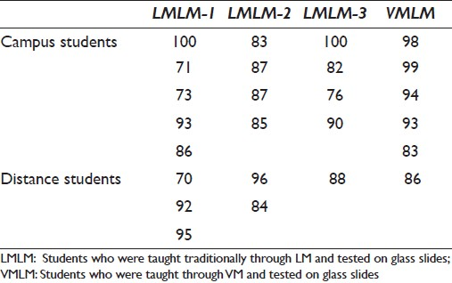
After screening the unknown virtual images of the three body sites, the students were given a final screening and interpretation examination, consisting of 10 conventional glass slides [Table 4] from all three sites, breast (n = 3), thyroid (n = 3), and lymph node (n = 4). This was the first time the students had screened the conventional glass slides with LM from these three body sites. The glass slides used for the examination were the same slides from a variety of diagnostic categories that had been used for testing the past three years. The participant’s scores were compared with the scores of the previous three years of students (n = 18) who learned with LM and were testing using LM [Table 5].
Medians and the twenty-fifth and seventy-fifth percentiles were used to show the test results. Counts and percentages were used to display responses from the survey. The Kruskal-Wallis test was used to compare the examination results from three years of prior testing, using glass slides. The Mann-Whitney test was used to compare the study participants’ glass slide examination scores, with the examination scores of students from the past three years. All tests were two-sided and a p-value < 0.05 was considered statistically significant. The analysis was conducted using the SPSS software. At the end of the study the students were sent a link to an online survey website ( www.surveymonkey.com) and asked to respond anonymously and voluntarily to a series of questions, in which they were to answer, (a) strongly agree, (b) agree, (c) neutral, (d) disagree or (e) strongly disagree. The survey also had an open comment section for the students to give any additional suggestions, to explain / justify their answers and to add any other comments not mentioned in the series of questions.
RESULTS
Objective variables
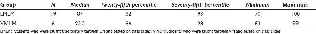
The final glass slide screening test scores of the participating students who were taught through VM and tested on glass slides (VMLM group) were compared with the last three classes of students who were taught traditionally through LM and tested on glass slides (LMLM group). There was no statistically significant difference in the median test scores of the last three classes of students (LMLM group, p = 0.97). Comparison of the median test scores from the VMLM and LMLM group indicated no statistically significant difference (p = 0.20). The median test score for the VMLM group was 93.5 (IQR = 12) and for the LMLM group it was 87 (IQR = 11) [Table 6].
Subjective variables
Annotations and method of learning
Students’ opinions about the annotations were completely positive. Two students indicated in the open comment section that the annotations were well presented and using VM made it easier to take notes for studying than when sitting at the multi-head microscope for a live teaching session. Four out of six students agreed that it was easy to apply the experience gained by VM in interpreting the traditional glass slides. Group discussions among students were found to be good and useful for half of the students. When their opinion was asked on whether they obtained optimized information when learning cytology through VM, the responses were diverse: two of the students agreed, one had a neutral opinion, and the other three disagreed.
Quality and navigation of virtual images
Three students had neutral opinions on switching the magnification of the virtual images from one to another; however, two students felt it was easy. Although the students did fairly well in diagnosing the unknown virtual images, most of their (n = 4) responses in the survey for the statement, ‘The resolution of the images was sufficient enough to give the diagnosis’ was neutral. Half of the students thought that 3D images would have been better. Overall, the students were not pleased about the time it took to download the virtual images and switch from one slide / image to another within the website.
Ease of virtual image screening
There was a range of responses on the statement about comparing the stress on students’ eyes in using VM and LM. Even though two students agreed to the statement that the VM experience was less stressful to the body than sitting behind a microscope, four responses were neutral. It was also noted that they did not consider the VM experience enjoyable and fun.
Five out of six students disagreed that screening virtual slides / images was easy. Some of the comments on this issue were, “it took a long time to screen the slides because every move required it to reload, which included changing magnification,” “screening virtual slides takes a long time and makes it hard to know what you have and haven't screened,” “light microscopy is faster and easier than the virtual microscopy.”
DISCUSSION
The objectives of this study were: (1) To determine that learning through VM could be applied effectively to glass slide screening using LM, and (2) to evaluate students’ opinions of using virtual slides compared to glass slides.
As the results indicated, the main objectives of this study were supported by the findings, which demonstrate that learning through VM could be successfully applied to glass slide screening using LM, with our six participants. The responses regarding the experience of using virtual slides compared to glass slides, however, were diverse. Even though the students indicated they did not want to replace LM with VM for learning cytology, they gave positive comments about their experiences.
One of the main aspects of VM that all the students appreciated was the annotations of the teaching slides. All students agreed that the annotations were more helpful than taking notes during the multi-head microscope sessions with LM; this was also mentioned twice in the open comment area. Four of the six students (66.6%) agreed that applying the knowledge and experience gained by learning cytology through VM on glass slide screening, using LM, was easy.
The participants in this study faced some technical issues that made their VM screening experience suboptimal. Even though only a portion of the slides were scanned, the image file sizes (average file size - 426.52 MB) were found to be too large, and it took very long to download each image and to switch between images. Some of these technical issues might be explained by the fact that students used their own computers to access images for this study. Therefore, all computer systems and connections to the internet were different, which might have affected the resolution and time to download the images. The students were also discouraged about the time required for them to screen the virtual images. They reported that more time was spent screening the virtual images compared to screening glass slides. Actual screening times were not collected for this project, so it was difficult to assess whether the participants only perceived that screening the virtual images took longer, or whether their statements were accurate. If they were in fact accurate, the authors think this could be due to a learning curve with the virtual image software and over time the screening time for virtual images (VM) would decrease, just as the screening time with glass slides (LM) has decreased. Another possible reason for the students’ perceived increase in screening time could be due to the presence of 3D cell clusters. These clusters might have required more time to read and interpret the cellular morphology. Taking this into account and also the survey responses of students who mentioned they would have preferred 3D images, we are currently investigating the optimal scanning parameters for cytology specimens.[31]
The software used to screen the virtual images presented another issue. The students were disappointed that the software did not allow them to mark the cells of interest, to enable them to go over them with the faculty member or fellow student at a later time. One of the students mentioned that many of the areas of the virtual images might have been missed while screening, since a straight line could not be followed as was done with glass slide screening. Hence, it was suggested that the arrow keys would have been a better option to screen the virtual images. The cytotechnology program has since switched software for the virtual images and screening with the arrow keys is now possible.
Another major concern of the students was lack of faculty member's interaction during the study period. The students strongly believed that the faculty member′s guidance and interaction would have better improved their practice of virtual slide screening. Faculty interaction was not available during this study in order to evaluate whether the virtual images could stand on their own as a primary method of teaching the students morphological criteria. In a typical cytotechnology teaching program, a faculty member would always be available to the students for questions and clarification. Perhaps the lack of faculty instruction and the software issues played a role in the majority of students (four out of six or 66.6%) responding that they disagreed that VM was fun and enjoyable.
The authors were surprised that half of the students stated that VM did not enhance their ability to learn cytology in spite of their better performance on the final glass slide screening when compared to the LMLM group. Also, only half of the students stated that being able to use VM outside the classroom (anytime, anywhere) gave them more time to study the images. The authors presumed that the students would spend more time looking at the images as opposed to glass slides, as they would not have to stay in the classroom to use LM. The amount of time spent previously looking at glass slides for other body sites and the amount of time spent looking at virtual images were not documented in this study; therefore, it is difficult to know whether the students actually spent longer studying one method more than the other.
Overall, the students considered VM a great tool for teaching and studying along with LM, but not for daily practice screening. Preference was given to LM, as the participants perceived the glass slide screening as easier, requiring less time to screen, and one could dot the cells of interest to discuss with a faculty member. It is important to mention the students′ previous exposure and experience with LM and faculty interaction. The students participating in this project were beginning their second semester in the cytotechnology program. During their first semester, they became accustomed to using LM with glass slides by screening them on a daily basis; therefore, their experience with VM was considered something new. The authors think the primary reason for the overall preference to LM was the students'; lack of training and experience with VM screening. In addition, our study was conducted with a limited number of students, for a three-week period, during the middle of the students′ academic course. The students had interaction with faculty members during the entire first semester while screening glass slides, so learning with VM annotations might have been considered different than their previous experience.
Future studies will need to be conducted to determine if students learning with VM from the first day of class have a different opinion about VM than in this study. It is believed that just like LM, VM takes practice. If they learn by using VM at the beginning of the course, they will attain proficiency in VM screening, which could make the learning experience with VM similar to LM screening.
CONCLUSION AND SUMMARY
The faculty and the students of the UNMC Cytotechnology program consider VM as a valuable teaching tool. Our study demonstrated that learning cytology through VM could be applied to glass slide screening using LM. However, most students preferred learning through LM and using virtual microscopy as an additional study tool.
COMPETING INTEREST STATEMENT BY ALL AUTHORS
The author(s) declare that they do not have competing commercial interests.
AUTHORSHIP STATEMENT BY ALL AUTHORS
All authors of this article declare that they qualify for authorship as defined by ICMJE http://www.icmje.org/#author. Each author has participated sufficiently in the study and take public responsibility for appropriate portions of the content of this article. Each author acknowledges that this final version has been read and approved by them.
ETHICS STATEMENT BY ALL AUTHORS
This study was conducted with approval from the Institutional Review Board (IRB) of all the institutions associated with this study. The authors take the responsibility of maintaining relevant documentation in this respect.
EDITORIAL / PEER-REVIEW STATEMENT
To ensure the integrity and highest quality of CytoJournal publications, the review process of this manuscript was conducted under a double blind model (authors are blinded for reviewers and vice versa) through automatic online system.
ACKNOWLEDGMENTS
This project was funded by the School of Allied Health, UNMC. The authors are very thankful to David Wert, Tissue Sciences Facility Supervisor, Department of Pathology and Microbiology, UNMC, for his expertise in the scanning of glass slides and producing virtual images.
Available FREE in open access from: http://www.cytojournal.com/text.asp?2012/9/1/12/95827.
REFERENCES
- Reading virtual slide using web viewers: results of subjective experience with three different solutions. Diagn Pathol. 2008;3(Suppl 1):S23.
- [Google Scholar]
- Digital pathology: exploring its applications in diagnostic surgical pathology practice. Pathology. 2010;42:512-8.
- [Google Scholar]
- Review of the current state of whole slide imaging in pathology. J Pathol Inform. 2011;2:36.
- [Google Scholar]
- Transition of a dental histology course from light to virtual microscopy. J Dent Educ. 2009;73:1213-21.
- [Google Scholar]
- Teaching medical histology at the University of South Carolina School of Medicine: Transition to virtual slides and virtual microscopes. Anat Rec B New Anat. 2003;275:196-206.
- [Google Scholar]
- Implementing digital technology to enhance student learning of pathology. Eur J Dent Educ. 2009;13:172-8.
- [Google Scholar]
- Introduction and evaluation of virtual microscopy in teaching veterinary cytopathology. J Vet Med Educ. 2007;34:437-44.
- [Google Scholar]
- Comparison of a virtual microscope laboratory to a regular microscope laboratory for teaching histology. Anat Rec. 2001;265:10-4.
- [Google Scholar]
- Integrated approach to teaching and testing in histology with real and virtual imaging. Anat Rec. 2002;269:107-12.
- [Google Scholar]
- Teaching histology to first-year veterinary science students using virtual microscopy and traditional microscopy: a comparison of student responses. J Vet Med Educ. 2007;34:177-82.
- [Google Scholar]
- Quantitative and qualitative changes in teaching histology by means of virtual microscopy in an introductory course in human anatomy. Anat Sci Educ. 2009;2:218-26.
- [Google Scholar]
- Teaching pathology via online digital microscopy: positive learning outcomes for rurally based medical students. Aust J Rural Health. 2011;19:45-51.
- [Google Scholar]
- Telecytology: diagnostic accuracy in cervical-vaginal smears. Am J Clin Pathol. 1996;105:599-603.
- [Google Scholar]
- Diagnostic concordance of telecytology and conventional cytology for evaluating breast aspirates. Acta Cytol. 1998;42:663-7.
- [Google Scholar]
- Telecytologic diagnosis of breast fine needle aspiration biopsies.Intraobserver concordance. Acta Cytol. 2000;44:175-80.
- [Google Scholar]
- Telecytology of fine-needle aspiration biopsies of the pancreas: a study of well-differentiated adenocarcinoma and chronic pancreatitis with atypical epithelial repair changes. Diagn Cytopathol. 2003;28:147-52.
- [Google Scholar]
- Telecytology in Hokkaido Island, Japan: results of primary telecytodiagnosis of routine cases. Cytopathology. 2004;15:221-7.
- [Google Scholar]
- An analysis of 46 static telecytology cases over a period of two years. J Telemed Telecare. 2006;12:311-4.
- [Google Scholar]
- Internet-based gynecologic telecytology with remote automated image selection: results of a first-phase developmental trial. Am J Clin Pathol. 2008;129:686-96.
- [Google Scholar]
- Telecytology: a tool for quality assessment and improvement in the evaluation of thyroid fine-needle aspiration specimens. Telemed J E Health. 2009;15:713-7.
- [Google Scholar]
- Use of telecytology for the immediate assessment of CT guided and endoscopic FNA cytology: Diagnostic accuracy, advantages, and pitfalls. Diagn Cytopathol 2010 [In Press]
- [Google Scholar]
- Dynamic telecytopathology of on site rapid cytology diagnoses for pancreatic carcinoma. CytoJournal. 2006;3:27.
- [Google Scholar]
- The impact of digital imaging in the field of cytopathology. CytoJournal. 2009;6:6.
- [Google Scholar]
- Telecytology for immediate assessment of endoscopic ultrasound-guided fine needle aspiration: Improved pathologist efficiency, and a valuable educational tool. CytoJournal. 2011;8:16.
- [Google Scholar]
- Virtual microscopy: an educator';s tool for the enhancement of cytotechnology students' locator skills. Diagn Cytopathol. 2008;36:363-8.
- [Google Scholar]
- Virtual microscopy as a tool for proficiency testing in cytopathology: a model using multiple digital images of Papanicolaou tests. Arch Pathol Lab Med. 2003;127:1320-4.
- [Google Scholar]
- Comparison of cytology proficiency testing: glass slides vs.virtual slides. Acta Cytol. 2004;48:788-94.
- [Google Scholar]
- Virtual microscopy for cytology proficiency testing: are we there yet? Cancer. 2007;111:203-9.
- [Google Scholar]
- The optimal z-axis interval and focal planes to digitize 3-D gynecological SurePath® glass slides: Initial findings. CytoJournal. 2011;8:16.
- [Google Scholar]


